 Movie
Movie Controller


+ Open data

- Basic information
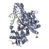
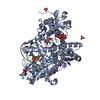
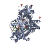
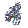
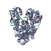
| Entry | Database: PDB / ID: 1nk0 | |||||||||
|---|---|---|---|---|---|---|---|---|---|---|
| Title | ADENINE-GUANINE MISMATCH AT THE POLYMERASE ACTIVE SITE | |||||||||
 Components Components |
| |||||||||
 Keywords Keywords | TRANSFERASE/DNA / DNA POLYMERASE I / DNA REPLICATION / KLENOW FRAGMENT / PROTEIN-DNA COMPLEX / DNA MISMATCH / TRANSFERASE-DNA COMPLEX | |||||||||
| Function / homology |  Function and homology information Function and homology information5'-3' exonuclease activity / 3'-5' exonuclease activity / DNA-templated DNA replication / double-strand break repair / DNA-directed DNA polymerase / DNA-directed DNA polymerase activity / DNA binding Similarity search - Function | |||||||||
| Biological species |   Geobacillus stearothermophilus (bacteria) Geobacillus stearothermophilus (bacteria) | |||||||||
| Method |  X-RAY DIFFRACTION / SYNCHROTRON / MOLECULAR REPLACEMENT / Resolution: 1.7 Å X-RAY DIFFRACTION / SYNCHROTRON / MOLECULAR REPLACEMENT / Resolution: 1.7 Å | |||||||||
 Authors Authors | Johnson, S.J. / Beese, L.S. | |||||||||
 Citation Citation | Journal: Cell(Cambridge,Mass.) / Year: 2004 Title: Structures of mismatch replication errors observed in a DNA polymerase. Authors: Johnson, S.J. / Beese, L.S. #1: Journal: Proc.Natl.Acad.Sci.USA / Year: 2003Title: Processive DNA synthesis observed in a polymerase crystal suggests a mechanism for the prevention of frameshift mutations. Authors: Johnson, S.J. / Taylor, J.S. / Beese, L.S. #2: Journal: Nature / Year: 1998Title: Visualizing DNA Replication in a Catalytically Active Bacillus DNA Polymerase Crystal Authors: Kiefer, J.R. / Mao, C. / Braman, J.C. / Beese, L.S. #3: Journal: Structure / Year: 1997Title: Crystal Structure of a Thermostable Bacillus DNA Polymerase I Large Fragment at 2.1 A Resolution Authors: Kiefer, J.R. / Mao, C. / Hansen, C.J. / Basehore, S.L. / Hogrefe, H.H. / Braman, J.C. / Beese, L.S. | |||||||||
| History |
|
- Structure visualization
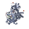
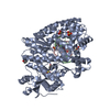
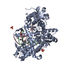
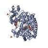
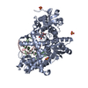
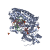
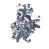
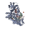
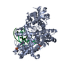
| Structure viewer | Molecule: MolmilJmol/JSmol |
|---|
- Downloads & links
-Download
| PDBx/mmCIF format | 1nk0.cif.gz | 163.2 KB | Display | PDBx/mmCIF format |
|---|---|---|---|---|
| PDB format | pdb1nk0.ent.gz | 119.7 KB | Display | PDB format |
| PDBx/mmJSON format | 1nk0.json.gz | Tree view | PDBx/mmJSON format | |
| Others |  Other downloads Other downloads |
-Validation report
| Arichive directory | https://data.pdbj.org/pub/pdb/validation_reports/nk/1nk0ftp://data.pdbj.org/pub/pdb/validation_reports/nk/1nk0 | HTTPS FTP |
|---|
-Related structure data
| Related structure data |  1njwC  1njxC 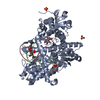 1njyC  1njzC  1nk4C  1nk5C 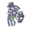 1nk6C 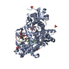 1nk7C 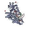 1nk8C 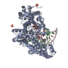 1nk9C 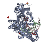 1nkbC  1nkcC  1nkeC  2bdpS C: citing same article ( S: Starting model for refinement |
|---|---|
| Similar structure data |


-Links
 PDBj
PDBj


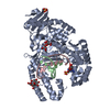
- Assembly
| Deposited unit | 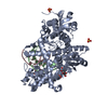
| ||||||||
|---|---|---|---|---|---|---|---|---|---|
| 1 |
| ||||||||
| Unit cell |
| ||||||||
| Details | Exists as a monomer. One molecule per asymmetric unit. |
-Components
-DNA chain , 2 types, 2 molecules BC
| #1: DNA chain | Mass: 3367.225 Da / Num. of mol.: 1 / Source method: obtained synthetically |
|---|---|
| #2: DNA chain | Mass: 4600.983 Da / Num. of mol.: 1 / Source method: obtained synthetically |
-Protein / Sugars , 2 types, 2 molecules A
| #3: Protein | Mass: 66172.844 Da / Num. of mol.: 1 Fragment: BACILLUS FRAGMENT (ANALOGOUS TO THE E. COLI KLENOW FRAGMENT) Source method: isolated from a genetically manipulated source Source: (gene. exp.) Geobacillus stearothermophilus (bacteria)Plasmid: PET-30A(+) / Production host: |
|---|---|
| #4: Polysaccharide | beta-D-fructofuranose-(2-1)-alpha-D-glucopyranose / sucrose  Source method: isolated from a genetically manipulated source Details: oligosaccharide with reducing-end-to-reducing-end glycosidic bond References: sucrose |
-Non-polymers , 3 types, 663 molecules 


| #5: Chemical | ChemComp-MES /  Mass: 195.237 Da / Num. of mol.: 1 / Source method: obtained synthetically / Formula: C6H13NO4S / Comment: pH buffer*YM Mass: 195.237 Da / Num. of mol.: 1 / Source method: obtained synthetically / Formula: C6H13NO4S / Comment: pH buffer*YM | ||
|---|---|---|---|
| #6: Chemical | ChemComp-SO4 /  Mass: 96.063 Da / Num. of mol.: 4 / Source method: obtained synthetically / Formula: SO4 Mass: 96.063 Da / Num. of mol.: 4 / Source method: obtained synthetically / Formula: SO4#7: Water | ChemComp-HOH / | Mass: 18.015 Da / Num. of mol.: 658 / Source method: isolated from a natural source / Formula: H2O |
-Details
| Compound details | THE POLYMERASE-DNA COMPLEX ADOPTS A DISTORTED OPEN CONFORMATION WITH AN ADENINE-GUANINE MISMATCH ...THE POLYMERASE |
|---|
-Experimental details
-Experiment
| Experiment | Method: X-RAY DIFFRACTION / Number of used crystals: 1 |
|---|
- Sample preparation
| Crystal | Density Matthews: 2.65 Å3/Da / Density % sol: 53.17 % | |||||||||||||||||||||||||||||||||||||||||||||||||||||||||||||||
|---|---|---|---|---|---|---|---|---|---|---|---|---|---|---|---|---|---|---|---|---|---|---|---|---|---|---|---|---|---|---|---|---|---|---|---|---|---|---|---|---|---|---|---|---|---|---|---|---|---|---|---|---|---|---|---|---|---|---|---|---|---|---|---|---|
| Crystal grow | Temperature: 290 K / Method: vapor diffusion, hanging drop / pH: 5.8 Details: AMMONIUM SULFATE, MAGNESIUM SULFATE, MPD, MES, pH 5.80, VAPOR DIFFUSION, HANGING DROP, temperature 290K | |||||||||||||||||||||||||||||||||||||||||||||||||||||||||||||||
| Components of the solutions |
| |||||||||||||||||||||||||||||||||||||||||||||||||||||||||||||||
| Crystal grow | *PLUS pH: 5.8 / Method: vapor diffusion, hanging dropDetails: Johnson, S.J., (2003) Proc.Natl.Acad.Sci.USA, 100, 3895. | |||||||||||||||||||||||||||||||||||||||||||||||||||||||||||||||
| Components of the solutions | *PLUS
|
-Data collection
| Diffraction |
| |||||||||
|---|---|---|---|---|---|---|---|---|---|---|
| Diffraction source | Source: SYNCHROTRON / Site: NSLS  / Beamline: X12B / Wavelength: 1.0001 Å / Beamline: X12B / Wavelength: 1.0001 Å | |||||||||
| Detector | Type: ADSC QUANTUM 4 / Detector: CCD / Date: Oct 13, 2000 | |||||||||
| Radiation | Monochromator: Si CRYSTALS / Protocol: SINGLE WAVELENGTH / Monochromatic (M) / Laue (L): M / Scattering type: x-ray | |||||||||
| Radiation wavelength | Wavelength: 1.0001 Å / Relative weight: 1 | |||||||||
| Reflection | Resolution: 1.7→50 Å / Num. all: 94629 / Num. obs: 94629 / % possible obs: 98.2 % / Observed criterion σ(F): -3 / Observed criterion σ(I): -3 / Redundancy: 3.1 % / Biso Wilson estimate: 15.3 Å2 / Rsym value: 0.047 / Net I/σ(I): 20.2 | |||||||||
| Reflection shell | Resolution: 1.7→1.76 Å / Redundancy: 2.5 % / Mean I/σ(I) obs: 3.3 / Num. unique all: 8835 / Rsym value: 0.26 / % possible all: 92.9 | |||||||||
| Reflection | *PLUS Num. measured all: 292563 / Rmerge(I) obs: 0.047 | |||||||||
| Reflection shell | *PLUS Highest resolution: 1.7 Å / Rmerge(I) obs: 0.26 |
- Processing
| Software |
| ||||||||||||||||||||||||||||||||||||||||||||||||||||||||||||||||||||||||||||||||
|---|---|---|---|---|---|---|---|---|---|---|---|---|---|---|---|---|---|---|---|---|---|---|---|---|---|---|---|---|---|---|---|---|---|---|---|---|---|---|---|---|---|---|---|---|---|---|---|---|---|---|---|---|---|---|---|---|---|---|---|---|---|---|---|---|---|---|---|---|---|---|---|---|---|---|---|---|---|---|---|---|---|
| Refinement | Method to determine structure: MOLECULAR REPLACEMENT Starting model: PDB ENTRY 2BDP Resolution: 1.7→28.32 Å / Rfactor Rfree error: 0.003 / Data cutoff high absF: 2153708.32 / Data cutoff high rms absF: 2153708.32 / Data cutoff low absF: 0 / Isotropic thermal model: RESTRAINED / Cross valid method: THROUGHOUT / σ(F): 2 / Stereochemistry target values: Engh & Huber Details: THE BACILLUS POLYMERASE WAS CO- CRYSTALLIZED WITH A DNA CONTAINING AN 11 BASE PAIR DUPLEX AND A 4 BASE 5' TEMPLATE OVERHANG. THE LAST THREE BASES IN THE OVERHANG ARE DISORDERED AND ARE NOT ...Details: THE BACILLUS POLYMERASE WAS CO- CRYSTALLIZED WITH A DNA CONTAINING AN 11 BASE PAIR DUPLEX AND A 4 BASE 5' TEMPLATE OVERHANG. THE LAST THREE BASES IN THE OVERHANG ARE DISORDERED AND ARE NOT INCLUDED IN THE MODEL. AN ADENINE-GUANINE MISMATCH IS LOCATED AT THE 3' PRIMER TERMINUS OF THE DNA DUPLEX. ELECTRON DENSITY WAS OBSERVED FOR ALL PROTEIN SIDE CHAINS EXCEPT LYSINE 298, WHICH WAS MODELLED TO THE BETA CARBON.
| ||||||||||||||||||||||||||||||||||||||||||||||||||||||||||||||||||||||||||||||||
| Solvent computation | Solvent model: FLAT MODEL / Bsol: 44.3887 Å2 / ksol: 0.378445 e/Å3 | ||||||||||||||||||||||||||||||||||||||||||||||||||||||||||||||||||||||||||||||||
| Displacement parameters | Biso mean: 20.8 Å2
| ||||||||||||||||||||||||||||||||||||||||||||||||||||||||||||||||||||||||||||||||
| Refine analyze |
| ||||||||||||||||||||||||||||||||||||||||||||||||||||||||||||||||||||||||||||||||
| Refinement step | Cycle: LAST / Resolution: 1.7→28.32 Å
| ||||||||||||||||||||||||||||||||||||||||||||||||||||||||||||||||||||||||||||||||
| Refine LS restraints |
| ||||||||||||||||||||||||||||||||||||||||||||||||||||||||||||||||||||||||||||||||
| LS refinement shell | Resolution: 1.7→1.76 Å / Rfactor Rfree error: 0.012 / Total num. of bins used: 10
| ||||||||||||||||||||||||||||||||||||||||||||||||||||||||||||||||||||||||||||||||
| Xplor file |
| ||||||||||||||||||||||||||||||||||||||||||||||||||||||||||||||||||||||||||||||||
| Refinement | *PLUS Highest resolution: 1.7 Å / Lowest resolution: 50 Å / Rfactor Rfree: 0.213 / Rfactor Rwork: 0.193 / % reflection Rfree: 5 % | ||||||||||||||||||||||||||||||||||||||||||||||||||||||||||||||||||||||||||||||||
| Solvent computation | *PLUS | ||||||||||||||||||||||||||||||||||||||||||||||||||||||||||||||||||||||||||||||||
| Displacement parameters | *PLUS | ||||||||||||||||||||||||||||||||||||||||||||||||||||||||||||||||||||||||||||||||
| Refine LS restraints | *PLUS
| ||||||||||||||||||||||||||||||||||||||||||||||||||||||||||||||||||||||||||||||||
| LS refinement shell | *PLUS Rfactor Rfree: 0.247 / Rfactor Rwork: 0.246 |
