 Movie
Movie Controller

[English] 日本語
 Yorodumi
- PDB-2he2: Crystal structure of the 3rd PDZ domain of human discs large homo... -
+ Open data

- Basic information
| Entry | Database: PDB / ID: 2he2 | ||||||
|---|---|---|---|---|---|---|---|
| Title | Crystal structure of the 3rd PDZ domain of human discs large homologue 2, DLG2 | ||||||
 Components Components | Discs large homolog 2 | ||||||
 Keywords Keywords | SIGNALING PROTEIN / DLG2 / PDZ / PDZ domain / signal transduction / Structural Genomics / Structural Genomics Consortium / SGC | ||||||
| Function / homology |  Function and homology information Function and homology informationretrograde axonal protein transport / anterograde axonal protein transport / GMP kinase activity / receptor localization to synapse / protein localization to synapse / establishment or maintenance of epithelial cell apical/basal polarity / juxtaparanode region of axon / cellular response to potassium ion / Assembly and cell surface presentation of NMDA receptors / Neurexins and neuroligins ...retrograde axonal protein transport / anterograde axonal protein transport / GMP kinase activity / receptor localization to synapse / protein localization to synapse / establishment or maintenance of epithelial cell apical/basal polarity / juxtaparanode region of axon / cellular response to potassium ion / Assembly and cell surface presentation of NMDA receptors / Neurexins and neuroligins / receptor clustering / Negative regulation of NMDA receptor-mediated neuronal transmission / Unblocking of NMDA receptors, glutamate binding and activation / Long-term potentiation / regulation of postsynaptic membrane neurotransmitter receptor levels / ionotropic glutamate receptor binding / axon cytoplasm / Ras activation upon Ca2+ influx through NMDA receptor / adherens junction / neuromuscular junction / cell-cell adhesion / postsynaptic density membrane / kinase binding / nervous system development / RAF/MAP kinase cascade / perikaryon / basolateral plasma membrane / chemical synaptic transmission / neuron projection / postsynaptic density / protein kinase binding / membrane / plasma membrane / cytosol Similarity search - Function | ||||||
| Biological species |  Homo sapiens (human) Homo sapiens (human) | ||||||
| Method |  X-RAY DIFFRACTION / SYNCHROTRON / MOLECULAR REPLACEMENT / Resolution: 1.5 Å X-RAY DIFFRACTION / SYNCHROTRON / MOLECULAR REPLACEMENT / Resolution: 1.5 Å | ||||||
 Authors Authors | Turnbull, A.P. / Phillips, C. / Berridge, G. / Savitsky, P. / Smee, C.E.A. / Papagrigoriou, E. / Debreczeni, J. / Gorrec, F. / Elkins, J.M. / von Delft, F. ...Turnbull, A.P. / Phillips, C. / Berridge, G. / Savitsky, P. / Smee, C.E.A. / Papagrigoriou, E. / Debreczeni, J. / Gorrec, F. / Elkins, J.M. / von Delft, F. / Weigelt, J. / Edwards, A. / Arrowsmith, C. / Sundstrom, M. / Doyle, D.A. / Structural Genomics Consortium (SGC) | ||||||
 Citation Citation | Journal: Protein Sci. / Year: 2007 Title: Structure of PICK1 and other PDZ domains obtained with the help of self-binding C-terminal extensions. Authors: Elkins, J.M. / Papagrigoriou, E. / Berridge, G. / Yang, X. / Phillips, C. / Gileadi, C. / Savitsky, P. / Doyle, D.A. | ||||||
| History |
|
- Structure visualization
| Structure viewer | Molecule: MolmilJmol/JSmol |
|---|
- Downloads & links
-Download
| PDBx/mmCIF format | 2he2.cif.gz | 103.8 KB | Display | PDBx/mmCIF format |
|---|---|---|---|---|
| PDB format | pdb2he2.ent.gz | 80.3 KB | Display | PDB format |
| PDBx/mmJSON format | 2he2.json.gz | Tree view | PDBx/mmJSON format | |
| Others |  Other downloads Other downloads |
-Validation report
| Summary document | 2he2_validation.pdf.gz | 418.3 KB | Display | wwPDB validaton report |
|---|---|---|---|---|
| Full document | 2he2_full_validation.pdf.gz | 419 KB | Display | |
| Data in XML | 2he2_validation.xml.gz | 13.4 KB | Display | |
| Data in CIF | 2he2_validation.cif.gz | 20.6 KB | Display | |
| Arichive directory | https://data.pdbj.org/pub/pdb/validation_reports/he/2he2ftp://data.pdbj.org/pub/pdb/validation_reports/he/2he2 | HTTPS FTP |
-Related structure data
| Related structure data |  2bygC  2fcfC  2fneC 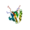 2gzvC 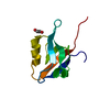 2he4C  2i1nC  2iwnC  2iwoC  2iwpC  2iwqC  1be9S  1bfeS  1tp3S  1tp5S S: Starting model for refinement C: citing same article ( |
|---|---|
| Similar structure data |


-Links
 PDBj
PDBj


- Assembly
| Deposited unit | 
| ||||||||
|---|---|---|---|---|---|---|---|---|---|
| 1 | 
| ||||||||
| 2 | 
| ||||||||
| Unit cell |
|
-Components
| #1: Protein | Mass: 10676.890 Da / Num. of mol.: 2 Source method: isolated from a genetically manipulated source Source: (gene. exp.) Homo sapiens (human) / Gene: DLG2 / Plasmid: pNIC28-Bsa4 / Production host:  #2: Water | ChemComp-HOH / |  Mass: 18.015 Da / Num. of mol.: 367 / Source method: isolated from a natural source / Formula: H2O Mass: 18.015 Da / Num. of mol.: 367 / Source method: isolated from a natural source / Formula: H2O |
|---|
-Experimental details
-Experiment
| Experiment | Method: X-RAY DIFFRACTION / Number of used crystals: 1 |
|---|
- Sample preparation
| Crystal | Density Matthews: 2.23 Å3/Da / Density % sol: 44.81 % |
|---|---|
| Crystal grow | Temperature: 293 K / Method: vapor diffusion, sitting drop Details: 25.5% PEG 3350; 0.17M (NH4)2SO4; 15% glycerol , VAPOR DIFFUSION, SITTING DROP, temperature 293K |
-Data collection
| Diffraction | Mean temperature: 100 K |
|---|---|
| Diffraction source | Source: SYNCHROTRON / Site: SLS  / Beamline: X10SA / Wavelength: 0.97646 Å / Beamline: X10SA / Wavelength: 0.97646 Å |
| Detector | Type: MARMOSAIC 225 mm CCD / Detector: CCD / Date: May 6, 2006 |
| Radiation | Monochromator: Si (111) / Protocol: SINGLE WAVELENGTH / Monochromatic (M) / Laue (L): M / Scattering type: x-ray |
| Radiation wavelength | Wavelength: 0.97646 Å / Relative weight: 1 |
| Reflection | Resolution: 1.5→50 Å / Num. obs: 30918 / % possible obs: 98.8 % / Observed criterion σ(F): 0 / Observed criterion σ(I): 0 |
| Reflection shell | Resolution: 1.5→1.55 Å / % possible all: 97.5 |
- Processing
| Software |
| ||||||||||||||||||||||||||||||||||||||||||||||||||||||||||||||||||||||||||||||||||||||||||||||||||||||||||||||||||||||||||||||||||||||||||||
|---|---|---|---|---|---|---|---|---|---|---|---|---|---|---|---|---|---|---|---|---|---|---|---|---|---|---|---|---|---|---|---|---|---|---|---|---|---|---|---|---|---|---|---|---|---|---|---|---|---|---|---|---|---|---|---|---|---|---|---|---|---|---|---|---|---|---|---|---|---|---|---|---|---|---|---|---|---|---|---|---|---|---|---|---|---|---|---|---|---|---|---|---|---|---|---|---|---|---|---|---|---|---|---|---|---|---|---|---|---|---|---|---|---|---|---|---|---|---|---|---|---|---|---|---|---|---|---|---|---|---|---|---|---|---|---|---|---|---|---|---|---|
| Refinement | Method to determine structure: MOLECULAR REPLACEMENT Starting model: Swissmodel based upon the coordinates of pdb entries 1TP5, 1TP3, 1BFE and 1BE9. Resolution: 1.5→41.13 Å / Cor.coef. Fo:Fc: 0.975 / Cor.coef. Fo:Fc free: 0.956 / SU B: 2.477 / SU ML: 0.043 / Cross valid method: THROUGHOUT / σ(F): 0 / ESU R: 0.077 / ESU R Free: 0.073 / Stereochemistry target values: MAXIMUM LIKELIHOOD / Details: HYDROGENS HAVE BEEN ADDED IN THE RIDING POSITIONS
| ||||||||||||||||||||||||||||||||||||||||||||||||||||||||||||||||||||||||||||||||||||||||||||||||||||||||||||||||||||||||||||||||||||||||||||
| Solvent computation | Ion probe radii: 0.8 Å / Shrinkage radii: 0.8 Å / VDW probe radii: 1.4 Å / Solvent model: MASK | ||||||||||||||||||||||||||||||||||||||||||||||||||||||||||||||||||||||||||||||||||||||||||||||||||||||||||||||||||||||||||||||||||||||||||||
| Displacement parameters | Biso mean: 9.045 Å2
| ||||||||||||||||||||||||||||||||||||||||||||||||||||||||||||||||||||||||||||||||||||||||||||||||||||||||||||||||||||||||||||||||||||||||||||
| Refinement step | Cycle: LAST / Resolution: 1.5→41.13 Å
| ||||||||||||||||||||||||||||||||||||||||||||||||||||||||||||||||||||||||||||||||||||||||||||||||||||||||||||||||||||||||||||||||||||||||||||
| Refine LS restraints |
| ||||||||||||||||||||||||||||||||||||||||||||||||||||||||||||||||||||||||||||||||||||||||||||||||||||||||||||||||||||||||||||||||||||||||||||
| LS refinement shell | Resolution: 1.5→1.539 Å / Total num. of bins used: 20
|
