 Movie
Movie Controller


+ Open data

- Basic information
| Entry | Database: PDB / ID: 1bfe | ||||||
|---|---|---|---|---|---|---|---|
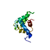
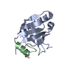
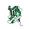
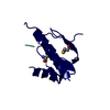
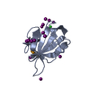
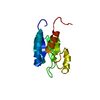
| Title | THE THIRD PDZ DOMAIN FROM THE SYNAPTIC PROTEIN PSD-95 | ||||||
 Components Components | PSD-95 | ||||||
 Keywords Keywords | PEPTIDE RECOGNITION / PROTEIN LOCALIZATION | ||||||
| Function / homology |  Function and homology information Function and homology informationRHO GTPases activate CIT / neuronal ion channel clustering / positive regulation of AMPA glutamate receptor clustering / P2Y1 nucleotide receptor binding / Neurexins and neuroligins / beta-1 adrenergic receptor binding / neuroligin family protein binding / regulation of grooming behavior / structural constituent of postsynaptic density / synaptic vesicle maturation ...RHO GTPases activate CIT / neuronal ion channel clustering / positive regulation of AMPA glutamate receptor clustering / P2Y1 nucleotide receptor binding / Neurexins and neuroligins / beta-1 adrenergic receptor binding / neuroligin family protein binding / regulation of grooming behavior / structural constituent of postsynaptic density / synaptic vesicle maturation / positive regulation of neuron projection arborization / receptor localization to synapse / cerebellar mossy fiber / LGI-ADAM interactions / protein localization to synapse / neuron spine / dendritic spine morphogenesis / Trafficking of AMPA receptors / proximal dendrite / negative regulation of receptor internalization / juxtaparanode region of axon / vocalization behavior / Activation of Ca-permeable Kainate Receptor / dendritic branch / acetylcholine receptor binding / positive regulation of dendrite morphogenesis / frizzled binding / cellular response to potassium ion / positive regulation of synapse assembly / dendritic spine organization / regulation of NMDA receptor activity / RAF/MAP kinase cascade / NMDA selective glutamate receptor signaling pathway / beta-2 adrenergic receptor binding / Synaptic adhesion-like molecules / neuromuscular process controlling balance / neurotransmitter receptor localization to postsynaptic specialization membrane / neuron projection terminus / cortical cytoskeleton / AMPA glutamate receptor clustering / locomotory exploration behavior / AMPA glutamate receptor complex / Unblocking of NMDA receptors, glutamate binding and activation / regulation of neuronal synaptic plasticity / glutamate receptor binding / social behavior / kinesin binding / D1 dopamine receptor binding / positive regulation of synaptic transmission / regulation of postsynaptic membrane neurotransmitter receptor levels / excitatory synapse / ionotropic glutamate receptor binding / dendrite cytoplasm / positive regulation of excitatory postsynaptic potential / cell periphery / synaptic membrane / PDZ domain binding / neuromuscular junction / establishment of protein localization / cell-cell adhesion / cerebral cortex development / regulation of long-term neuronal synaptic plasticity / postsynaptic density membrane / kinase binding / cell-cell junction / synaptic vesicle / cell junction / nervous system development / positive regulation of cytosolic calcium ion concentration / protein-containing complex assembly / scaffold protein binding / protein phosphatase binding / dendritic spine / chemical synaptic transmission / postsynaptic membrane / neuron projection / postsynaptic density / postsynapse / signaling receptor binding / synapse / dendrite / protein kinase binding / protein-containing complex binding / glutamatergic synapse / endoplasmic reticulum / membrane / plasma membrane / cytoplasm / cytosol Similarity search - Function | ||||||
| Biological species |  | ||||||
| Method |  X-RAY DIFFRACTION / MIR / Resolution: 2.3 Å X-RAY DIFFRACTION / MIR / Resolution: 2.3 Å | ||||||
 Authors Authors | Doyle, D.A. / Lee, A. / Lewis, J. / Kim, E. / Sheng, M. / Mackinnon, R. | ||||||
 Citation Citation | Journal: Cell(Cambridge,Mass.) / Year: 1996 Title: Crystal structures of a complexed and peptide-free membrane protein-binding domain: molecular basis of peptide recognition by PDZ. Authors: Doyle, D.A. / Lee, A. / Lewis, J. / Kim, E. / Sheng, M. / MacKinnon, R. #1: Journal: Nat.Struct.Biol. / Year: 1998Title: Crystal Structure of the Hcask Pdz Domain Reveals Structural Basis of Class II Pdz Domain Target Recognition Authors: Daniels, D.L. / Cohen, A.R. / Anderson, J.M. / Brunger, A.T. #2: Journal: Nature / Year: 1996Title: Crystal Structure of a Pdz Domain Authors: Cabral, M. / Petosa, C. / Sutcliffe, M.J. / Raza, S. / Byron, O. / Poy, F. / Marfatia, S.M. / Chishti, A.H. / Liddington, R.C. | ||||||
| History |
|
- Structure visualization
| Structure viewer | Molecule: MolmilJmol/JSmol |
|---|
- Downloads & links
-Download
| PDBx/mmCIF format | 1bfe.cif.gz | 34.2 KB | Display | PDBx/mmCIF format |
|---|---|---|---|---|
| PDB format | pdb1bfe.ent.gz | 24.2 KB | Display | PDB format |
| PDBx/mmJSON format | 1bfe.json.gz | Tree view | PDBx/mmJSON format | |
| Others |  Other downloads Other downloads |
-Validation report
| Arichive directory | https://data.pdbj.org/pub/pdb/validation_reports/bf/1bfeftp://data.pdbj.org/pub/pdb/validation_reports/bf/1bfe | HTTPS FTP |
|---|
-Related structure data


-Links
 PDBj
PDBj


- Assembly
| Deposited unit | 
| ||||||||||||
|---|---|---|---|---|---|---|---|---|---|---|---|---|---|
| 1 |
| ||||||||||||
| Unit cell |
| ||||||||||||
| Components on special symmetry positions |
|
-Components
| #1: Protein | Mass: 12750.126 Da / Num. of mol.: 1 / Fragment: THE THIRD PDZ DOMAIN OF PSD-95 Source method: isolated from a genetically manipulated source Source: (gene. exp.)  |
|---|---|
| #2: Water | ChemComp-HOH /  Mass: 18.015 Da / Num. of mol.: 111 / Source method: isolated from a natural source / Formula: H2O Mass: 18.015 Da / Num. of mol.: 111 / Source method: isolated from a natural source / Formula: H2O |
-Experimental details
-Experiment
| Experiment | Method: X-RAY DIFFRACTION / Number of used crystals: 1 |
|---|
- Sample preparation
| Crystal | Density Matthews: 2.32 Å3/Da / Density % sol: 46.95 % | ||||||||||||||||||||||||
|---|---|---|---|---|---|---|---|---|---|---|---|---|---|---|---|---|---|---|---|---|---|---|---|---|---|
| Crystal grow | pH: 7.5 / Details: 0.8 M SODIUM CITRATE, 0.1 M HEPES, PH 7.5 | ||||||||||||||||||||||||
| Crystal | *PLUS | ||||||||||||||||||||||||
| Crystal grow | *PLUS Method: vapor diffusion, sitting drop | ||||||||||||||||||||||||
| Components of the solutions | *PLUS
|
-Data collection
| Diffraction | Mean temperature: 100 K |
|---|---|
| Diffraction source | Source: ROTATING ANODE / Type: SIEMENS / Wavelength: 1.5418 |
| Detector | Type: MARRESEARCH / Detector: IMAGE PLATE / Date: Jan 1, 1996 / Details: COLLIMATOR |
| Radiation | Monochromator: NI FILTER / Monochromatic (M) / Laue (L): M / Scattering type: x-ray |
| Radiation wavelength | Wavelength: 1.5418 Å / Relative weight: 1 |
| Reflection | Resolution: 2.2→20 Å / Num. obs: 6421 / % possible obs: 96.6 % / Observed criterion σ(I): 2 / Rmerge(I) obs: 0.036 |
| Reflection shell | Resolution: 2.3→2.38 Å / Rmerge(I) obs: 0.166 / % possible all: 86.7 |
| Reflection | *PLUS Num. measured all: 38267 |
| Reflection shell | *PLUS % possible obs: 90.2 % |
- Processing
| Software |
| ||||||||||||
|---|---|---|---|---|---|---|---|---|---|---|---|---|---|
| Refinement | Method to determine structure: MIR / Resolution: 2.3→20 Å / Rfactor Rfree: 0.315 / Rfactor Rwork: 0.214 | ||||||||||||
| Refinement step | Cycle: LAST / Resolution: 2.3→20 Å
| ||||||||||||
| Software | *PLUS Name: X-PLOR / Classification: refinement | ||||||||||||
| Refinement | *PLUS Highest resolution: 2.3 Å / Rfactor obs: 0.214 / Rfactor Rfree: 0.315 / Lowest resolution: 6 Å / σ(F): 2 / % reflection Rfree: 10 % / Num. reflection obs: 5322 | ||||||||||||
| Solvent computation | *PLUS | ||||||||||||
| Displacement parameters | *PLUS | ||||||||||||
| Refine LS restraints | *PLUS
|
