 Movie
Movie Controller

[English] 日本語
 Yorodumi
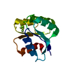
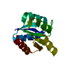
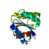
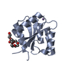
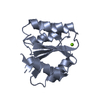
- PDB-2hdx: Crystal structure of the Src homology-2 domain of SH2-B in comple... -
+ Open data

- Basic information
| Entry | Database: PDB / ID: 2hdx | ||||||
|---|---|---|---|---|---|---|---|
| Title | Crystal structure of the Src homology-2 domain of SH2-B in complex with Jak2 pTyr813 phosphopeptide | ||||||
 Components Components |
| ||||||
 Keywords Keywords | SIGNALING PROTEIN / SH2 / Jak2 / phosphotyrosine / adapter protein | ||||||
| Function / homology |  Function and homology information Function and homology informationresponse to granulocyte macrophage colony-stimulating factor / IFNG signaling activates MAPKs / Regulation of IFNG signaling / positive regulation of cell activation / defense response to symbiont / Growth hormone receptor signaling / Signaling by Erythropoietin / Interleukin-4 and Interleukin-13 signaling / negative regulation of 3'-UTR-mediated mRNA stabilization / Interferon gamma signaling ...response to granulocyte macrophage colony-stimulating factor / IFNG signaling activates MAPKs / Regulation of IFNG signaling / positive regulation of cell activation / defense response to symbiont / Growth hormone receptor signaling / Signaling by Erythropoietin / Interleukin-4 and Interleukin-13 signaling / negative regulation of 3'-UTR-mediated mRNA stabilization / Interferon gamma signaling / Interleukin-20 family signaling / Interleukin-12 signaling / Signaling by CSF3 (G-CSF) / IL-6-type cytokine receptor ligand interactions / Erythropoietin activates RAS / MAPK3 (ERK1) activation / MAPK1 (ERK2) activation / Interleukin-23 signaling / Interleukin-27 signaling / negative regulation of growth hormone receptor signaling pathway / regulation of DNA biosynthetic process / Prolactin receptor signaling / Interleukin-6 signaling / Interleukin-35 Signalling / Signaling by SCF-KIT / RAF activation / tyrosine phosphorylation of STAT protein / T-helper 1 cell differentiation / Erythropoietin activates Phosphoinositide-3-kinase (PI3K) / Inactivation of CSF3 (G-CSF) signaling / Interleukin receptor SHC signaling / Cyclin D associated events in G1 / RAF/MAP kinase cascade / Factors involved in megakaryocyte development and platelet production / nuclear receptor-mediated mineralocorticoid signaling pathway / histone H3Y41 kinase activity / symbiont-induced defense-related programmed cell death / Interleukin-3, Interleukin-5 and GM-CSF signaling / positive regulation of growth hormone receptor signaling pathway / mammary gland epithelium development / positive regulation of growth factor dependent skeletal muscle satellite cell proliferation / regulation of postsynapse to nucleus signaling pathway / granulocyte macrophage colony-stimulating factor receptor complex / granulocyte-macrophage colony-stimulating factor signaling pathway / thrombopoietin-mediated signaling pathway / collagen-activated signaling pathway / activation of Janus kinase activity / response to interleukin-12 / interleukin-12 receptor binding / interleukin-5-mediated signaling pathway / positive regulation of leukocyte proliferation / type 1 angiotensin receptor binding / interleukin-12 receptor complex / interleukin-23 receptor complex / erythropoietin-mediated signaling pathway / post-embryonic hemopoiesis / myeloid cell differentiation / interleukin-23-mediated signaling pathway / positive regulation of T-helper 17 type immune response / interleukin-12-mediated signaling pathway / positive regulation of MHC class II biosynthetic process / positive regulation of NK T cell proliferation / transmembrane receptor protein tyrosine kinase adaptor activity / positive regulation of cell-substrate adhesion / positive regulation of platelet activation / interleukin-3-mediated signaling pathway / acetylcholine receptor binding / positive regulation of platelet aggregation / cellular response to interleukin-3 / growth hormone receptor binding / regulation of nitric oxide biosynthetic process / positive regulation of epithelial cell apoptotic process / response to hydroperoxide / axon regeneration / growth hormone receptor signaling pathway / intrinsic apoptotic signaling pathway in response to oxidative stress / positive regulation of tyrosine phosphorylation of STAT protein / extrinsic component of plasma membrane / lamellipodium assembly / negative regulation of cell-cell adhesion / extrinsic component of cytoplasmic side of plasma membrane / enzyme-linked receptor protein signaling pathway / peptide hormone receptor binding / platelet-derived growth factor receptor signaling pathway / positive regulation of interleukin-17 production / positive regulation of natural killer cell proliferation / signaling receptor activator activity / positive regulation of SMAD protein signal transduction / negative regulation of cardiac muscle cell apoptotic process / insulin receptor substrate binding / growth hormone receptor signaling pathway via JAK-STAT / response to tumor necrosis factor / cellular response to dexamethasone stimulus / type II interferon-mediated signaling pathway / cell surface receptor signaling pathway via JAK-STAT / phosphatidylinositol 3-kinase binding / positive regulation of vascular associated smooth muscle cell proliferation / extrinsic apoptotic signaling pathway / ruffle / negative regulation of protein localization to chromatin Similarity search - Function | ||||||
| Biological species |  | ||||||
| Method |  X-RAY DIFFRACTION / SYNCHROTRON / MOLECULAR REPLACEMENT / Resolution: 2.35 Å X-RAY DIFFRACTION / SYNCHROTRON / MOLECULAR REPLACEMENT / Resolution: 2.35 Å | ||||||
 Authors Authors | Hu, J. / Hubbard, S.R. | ||||||
 Citation Citation | Journal: J.Mol.Biol. / Year: 2006 Title: Structural Basis for Phosphotyrosine Recognition by the Src Homology-2 Domains of the Adapter Proteins SH2-B and APS. Authors: Hu, J. / Hubbard, S.R. | ||||||
| History |
|
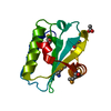
- Structure visualization
| Structure viewer | Molecule: MolmilJmol/JSmol |
|---|
- Downloads & links
-Download
| PDBx/mmCIF format | 2hdx.cif.gz | 148.8 KB | Display | PDBx/mmCIF format |
|---|---|---|---|---|
| PDB format | pdb2hdx.ent.gz | 118.9 KB | Display | PDB format |
| PDBx/mmJSON format | 2hdx.json.gz | Tree view | PDBx/mmJSON format | |
| Others |  Other downloads Other downloads |
-Validation report
| Arichive directory | https://data.pdbj.org/pub/pdb/validation_reports/hd/2hdxftp://data.pdbj.org/pub/pdb/validation_reports/hd/2hdx | HTTPS FTP |
|---|
-Related structure data
| Related structure data | 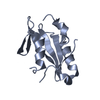 2hdvSC S: Starting model for refinement C: citing same article ( |
|---|---|
| Similar structure data |


-Links
 PDBj
PDBj


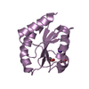
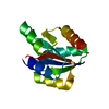
- Assembly
| Deposited unit | 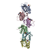
| ||||||||
|---|---|---|---|---|---|---|---|---|---|
| 1 | 
| ||||||||
| 2 | 
| ||||||||
| 3 | 
| ||||||||
| 4 | 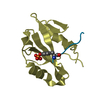
| ||||||||
| 5 | 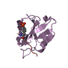
| ||||||||
| 6 | 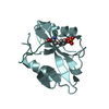
| ||||||||
| Unit cell |
| ||||||||
| Details | Six biological units pack in 12 5 screw axis |
-Components
| #1: Protein | Mass: 12275.992 Da / Num. of mol.: 6 / Fragment: SH2 (Residues: 499-607) / Mutation: E583A, E584A, W593H Source method: isolated from a genetically manipulated source Source: (gene. exp.)  #2: Protein/peptide | Mass: 1389.312 Da / Num. of mol.: 6 / Source method: obtained synthetically / Details: Sequence occurs naturally in Mus musculus (mouse). / References: UniProt: Q7TQD0, UniProt: Q62120*PLUS #3: Water | ChemComp-HOH / |  Mass: 18.015 Da / Num. of mol.: 320 / Source method: isolated from a natural source / Formula: H2O Mass: 18.015 Da / Num. of mol.: 320 / Source method: isolated from a natural source / Formula: H2OHas protein modification | Y | |
|---|
-Experimental details
-Experiment
| Experiment | Method: X-RAY DIFFRACTION / Number of used crystals: 1 |
|---|
- Sample preparation
| Crystal | Density Matthews: 2.39 Å3/Da / Density % sol: 48.56 % |
|---|---|
| Crystal grow | pH: 5.6 Details: 25% PEG 4000, 0.1 M sodium citrate, 0.3 M ammonium acetate., pH 5.6 |
-Data collection
| Diffraction | Mean temperature: 200 K |
|---|---|
| Diffraction source | Source: SYNCHROTRON / Site: NSLS  / Beamline: X4A / Wavelength: 1.5418 Å / Beamline: X4A / Wavelength: 1.5418 Å |
| Detector | Type: ADSC QUANTUM 4 / Detector: CCD / Date: May 1, 2005 |
| Radiation | Monochromator: GRAPHITE / Protocol: SINGLE WAVELENGTH / Monochromatic (M) / Laue (L): M / Scattering type: x-ray |
| Radiation wavelength | Wavelength: 1.5418 Å / Relative weight: 1 |
| Reflection | Resolution: 2.35→50 Å / Num. obs: 33327 / % possible obs: 98.1 % / Observed criterion σ(F): 1 / Observed criterion σ(I): 1 / Redundancy: 4.7 % / Rmerge(I) obs: 0.072 / Net I/σ(I): 16.1 |
| Reflection shell | Resolution: 2→2.07 Å / Redundancy: 3.3 % / Mean I/σ(I) obs: 10.2 / Rsym value: 0.103 / % possible all: 97.6 |
- Processing
| Software |
| ||||||||||||||||||||
|---|---|---|---|---|---|---|---|---|---|---|---|---|---|---|---|---|---|---|---|---|---|
| Refinement | Method to determine structure: MOLECULAR REPLACEMENT Starting model: PDB Entry: 2HDV Resolution: 2.35→30 Å / σ(F): 0 / Stereochemistry target values: Engh & Huber
| ||||||||||||||||||||
| Refinement step | Cycle: LAST / Resolution: 2.35→30 Å
| ||||||||||||||||||||
| Refine LS restraints |
|
