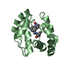
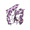
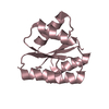
Crystal structure of DNA-binding response regulator, LuxR family, from Staphylococcus aureus
Components
Uncharacterized protein Q99UF4
Keywords
TRANSCRIPTION / STRUCTURAL GENOMICS / PSI-2 / PROTEIN STRUCTURE INITIATIVE / New York SGX Research Center for Structural Genomics / NYSGXRC / Q99UF4 / DNA-binding / Transcription regulation
Function / homology
Function and homology information
phosphorelay signal transduction system / transcription cis-regulatory region binding / regulation of DNA-templated transcription / DNA-templated transcription / DNA binding Similarity search - Function
Protocol: SINGLE WAVELENGTH / Monochromatic (M) / Laue (L): M / Scattering type: x-ray
Radiation wavelength
Wavelength: 0.979 Å / Relative weight: 1
Reflection
Redundancy: 6.4 % / Av σ(I) over netI: 10.2 / Number: 101117 / Rmerge(I) obs: 0.083 / Χ2: 1.31 / D res high: 2.04 Å / D res low: 50 Å / Num. obs: 15739 / % possible obs: 86.5
Diffraction reflection shell
Highest resolution (Å)
Lowest resolution (Å)
% possible obs (%)
ID
Rmerge(I) obs
Chi squared
Redundancy
4.39
50
99.5
1
0.046
1.655
8.2
3.49
4.39
100
1
0.065
2.077
8.3
3.05
3.49
100
1
0.097
1.449
8.4
2.77
3.05
100
1
0.15
1.135
8.3
2.57
2.77
99.9
1
0.226
0.91
7.8
2.42
2.57
99.7
1
0.279
0.832
6.1
2.3
2.42
97.4
1
0.321
0.83
4.4
2.2
2.3
88.3
1
0.344
0.936
3
2.11
2.2
56.9
1
0.383
0.756
2.1
2.04
2.11
22.9
1
0.436
0.84
1.6
Reflection
Resolution: 2.04→50 Å / Num. obs: 15739 / % possible obs: 86.5 % / Redundancy: 6.4 % / Rmerge(I) obs: 0.083 / Χ2: 1.311 / Net I/σ(I): 10.2
Reflection shell
Resolution (Å)
Redundancy (%)
Rmerge(I) obs
Num. unique all
Χ2
Diffraction-ID
% possible all
2.04-2.11
1.6
0.436
416
0.84
1
22.9
2.11-2.2
2.1
0.383
1036
0.756
1
56.9
2.2-2.3
3
0.344
1598
0.936
1
88.3
2.3-2.42
4.4
0.321
1760
0.83
1
97.4
2.42-2.57
6.1
0.279
1824
0.832
1
99.7
2.57-2.77
7.8
0.226
1804
0.91
1
99.9
2.77-3.05
8.3
0.15
1822
1.135
1
100
3.05-3.49
8.4
0.097
1820
1.449
1
100
3.49-4.39
8.3
0.065
1829
2.077
1
100
4.39-50
8.2
0.046
1830
1.655
1
99.5
-
Phasing
Phasing
Method: MAD
-
Processing
Software
Name
Version
Classification
NB
DENZO
datareduction
SCALEPACK
datascaling
SHELX
phasing
REFMAC
refinement
PDB_EXTRACT
3
dataextraction
CBASS
datacollection
SHELXD
phasing
Refinement
Method to determine structure: SAD / Resolution: 2.04→20 Å / Cor.coef. Fo:Fc: 0.969 / Cor.coef. Fo:Fc free: 0.938 / SU B: 8.077 / SU ML: 0.098 / TLS residual ADP flag: LIKELY RESIDUAL / Cross valid method: THROUGHOUT / σ(F): 0 / ESU R: 0.168 / ESU R Free: 0.159 / Stereochemistry target values: MAXIMUM LIKELIHOOD Details: The Friedel pairs were used in phasing. HYDROGENS HAVE BEEN ADDED IN THE RIDING POSITIONS
Rfactor
Num. reflection
% reflection
Selection details
Rfree
0.215
436
4.8 %
RANDOM
Rwork
0.165
-
-
-
obs
0.168
9087
89.86 %
-
Solvent computation
Ion probe radii: 0.8 Å / Shrinkage radii: 0.8 Å / VDW probe radii: 1.2 Å / Solvent model: BABINET MODEL WITH MASK
Displacement parameters
Biso mean: 38.829 Å2
Baniso -1
Baniso -2
Baniso -3
1-
0.06 Å2
0.03 Å2
0 Å2
2-
-
0.06 Å2
0 Å2
3-
-
-
-0.09 Å2
Refinement step
Cycle: LAST / Resolution: 2.04→20 Å
Protein
Nucleic acid
Ligand
Solvent
Total
Num. atoms
955
0
1
57
1013
Refine LS restraints
Refine-ID
Type
Dev ideal
Dev ideal target
Number
X-RAY DIFFRACTION
r_bond_refined_d
0.019
0.022
971
X-RAY DIFFRACTION
r_angle_refined_deg
1.661
1.985
1314
X-RAY DIFFRACTION
r_dihedral_angle_1_deg
6.49
5
121
X-RAY DIFFRACTION
r_dihedral_angle_2_deg
40.77
26.444
45
X-RAY DIFFRACTION
r_dihedral_angle_3_deg
17.342
15
193
X-RAY DIFFRACTION
r_dihedral_angle_4_deg
19.578
15
4
X-RAY DIFFRACTION
r_chiral_restr
0.114
0.2
161
X-RAY DIFFRACTION
r_gen_planes_refined
0.006
0.02
701
X-RAY DIFFRACTION
r_nbd_refined
0.212
0.2
468
X-RAY DIFFRACTION
r_nbtor_refined
0.31
0.2
691
X-RAY DIFFRACTION
r_xyhbond_nbd_refined
0.113
0.2
51
X-RAY DIFFRACTION
r_metal_ion_refined
0.153
0.2
2
X-RAY DIFFRACTION
r_symmetry_vdw_refined
0.258
0.2
34
X-RAY DIFFRACTION
r_symmetry_hbond_refined
0.386
0.2
11
X-RAY DIFFRACTION
r_mcbond_it
1.555
1.5
600
X-RAY DIFFRACTION
r_mcangle_it
5.504
20
981
X-RAY DIFFRACTION
r_scbond_it
11.587
20
374
X-RAY DIFFRACTION
r_scangle_it
6.446
4.5
332
LS refinement shell
Resolution: 2.04→2.09 Å / Total num. of bins used: 20
Rfactor
Num. reflection
% reflection
Rfree
0.215
8
-
Rwork
0.216
207
-
all
-
215
-
obs
-
-
29.99 %
Refinement TLS params.
Method: refined / Origin x: -4.5335 Å / Origin y: -26.7844 Å / Origin z: 4.5925 Å
11
12
13
21
22
23
31
32
33
T
-0.0936 Å2
0.0234 Å2
0.0116 Å2
-
-0.0413 Å2
0.0403 Å2
-
-
-0.0959 Å2
L
3.7113 °2
-1.2841 °2
0.9747 °2
-
2.4338 °2
-0.2646 °2
-
-
3.4012 °2
S
0.0061 Å °
0.1894 Å °
0.2113 Å °
-0.0191 Å °
-0.0257 Å °
0.0816 Å °
-0.2026 Å °
-0.1888 Å °
0.0196 Å °
+
About Yorodumi
-
News
-
Feb 9, 2022. New format data for meta-information of EMDB entries
New format data for meta-information of EMDB entries
Version 3 of the EMDB header file is now the official format.
The previous official version 1.9 will be removed from the archive.
In the structure databanks used in Yorodumi, some data are registered as the other names, "COVID-19 virus" and "2019-nCoV". Here are the details of the virus and the list of structure data.
Jan 31, 2019. EMDB accession codes are about to change! (news from PDBe EMDB page)
EMDB accession codes are about to change! (news from PDBe EMDB page)
The allocation of 4 digits for EMDB accession codes will soon come to an end. Whilst these codes will remain in use, new EMDB accession codes will include an additional digit and will expand incrementally as the available range of codes is exhausted. The current 4-digit format prefixed with “EMD-” (i.e. EMD-XXXX) will advance to a 5-digit format (i.e. EMD-XXXXX), and so on. It is currently estimated that the 4-digit codes will be depleted around Spring 2019, at which point the 5-digit format will come into force.
The EM Navigator/Yorodumi systems omit the EMD- prefix.
Related info.:Q: What is EMD? / ID/Accession-code notation in Yorodumi/EM Navigator
Yorodumi is a browser for structure data from EMDB, PDB, SASBDB, etc.
This page is also the successor to EM Navigator detail page, and also detail information page/front-end page for Omokage search.
The word "yorodu" (or yorozu) is an old Japanese word meaning "ten thousand". "mi" (miru) is to see.
Related info.:EMDB / PDB / SASBDB / Comparison of 3 databanks / Yorodumi Search / Aug 31, 2016. New EM Navigator & Yorodumi / Yorodumi Papers / Jmol/JSmol / Function and homology information / Changes in new EM Navigator and Yorodumi
 Movie
Movie Controller

 Yorodumi
 Open data
 Basic information
 Components
 Keywords
 Function and homology information
 Staphylococcus aureus (bacteria)
 X-RAY DIFFRACTION /
  Authors
 Citation
 Structure visualization
 Downloads & links
 Other downloads
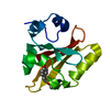


 PDBj
PDBj

 Assembly
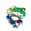

 Mass: 22.990 Da / Num. of mol.: 1 / Source method: obtained synthetically / Formula: Na
 Mass: 18.015 Da / Num. of mol.: 57 / Source method: isolated from a natural source / Formula: H2O
 Sample preparation
 / Beamline: X29A / Wavelength: 0.979 Å
 Processing