- PDB-3t6k: Crystal structure of a putative response regulator (Caur_3799) fr... -
+
Open data
ID or keywords:
Loading...
-
Basic information
Entry
Database: PDB / ID: 3t6k
Title
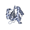
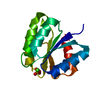
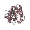
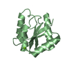
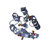
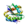
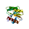
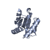
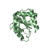
Crystal structure of a putative response regulator (Caur_3799) from Chloroflexus aurantiacus J-10-fl at 1.86 A resolution
Components
Response regulator receiver
Keywords
SIGNALING PROTEIN / Flavodoxin-like / Structural Genomics / Joint Center for Structural Genomics / JCSG / Protein Structure Initiative / PSI-BIOLOGY
Function / homology
Function and homology information
phosphorelay signal transduction system / cytoplasmic side of plasma membrane / ATP hydrolysis activity / ATP binding / cytosol Similarity search - Function
Mass: 18.015 Da / Num. of mol.: 367 / Source method: isolated from a natural source / Formula: H2O
Has protein modification
Y
Sequence details
THE CONSTRUCT (RESIDUES 1-135) WAS EXPRESSED WITH A PURIFICATION TAG MGSDKIHHHHHHENLYFQG. THE TAG ...THE CONSTRUCT (RESIDUES 1-135) WAS EXPRESSED WITH A PURIFICATION TAG MGSDKIHHHHHHENLYFQG. THE TAG WAS REMOVED WITH TEV PROTEASE LEAVING ONLY A GLYCINE (0) FOLLOWED BY THE TARGET SEQUENCE.
-
Experimental details
-
Experiment
Experiment
Method: X-RAY DIFFRACTION / Number of used crystals: 1
-
Sample preparation
Crystal
Density Matthews: 3.73 Å3/Da / Density % sol: 66.98 %
Crystal grow
Temperature: 277 K / Method: vapor diffusion, sitting drop Details: 20% polyethylene glycol 3350, 0.2M ammonium sulfate, VAPOR DIFFUSION,SITTING DROP,NANODROP, temperature 277K, VAPOR DIFFUSION, SITTING DROP
Type: MARMOSAIC 325 mm CCD / Detector: CCD / Date: Jul 20, 2011 Details: Flat mirror (vertical focusing); single crystal Si(111) bent monochromator (ho rizontal focusing)
Radiation
Monochromator: single crystal Si(111) bent / Protocol: MAD / Monochromatic (M) / Laue (L): M / Scattering type: x-ray
Radiation wavelength
ID
Wavelength (Å)
Relative weight
1
0.97916
1
2
0.91837
1
3
0.97876
1
Reflection
Resolution: 1.86→46.117 Å / Num. obs: 39465 / % possible obs: 99.9 % / Observed criterion σ(I): -3 / Biso Wilson estimate: 22.16 Å2 / Rmerge F obs: 0.145 / Rmerge(I) obs: 0.144 / Rrim(I) all: 0.15 / Net I/σ(I): 15.8 / Num. measured all: 472009
Reflection shell
Diffraction-ID: 1
Resolution (Å)
Highest resolution (Å)
Rmerge F obs
Rmerge(I) obs
Mean I/σ(I) obs
Num. measured obs
Num. possible
Num. unique obs
Rrim(I) all
% possible all
1.86-1.93
0.816
1.3
2.2
48857
4055
4052
1.358
99.9
1.93-2
0.54
0.924
3.1
42698
3535
3535
0.965
100
2-2.09
0.401
0.691
4.1
46341
3829
3828
0.722
100
2.09-2.2
0.29
0.499
5.6
47114
3889
3889
0.521
100
2.2-2.34
0.21
0.345
8
47967
3982
3971
0.36
99.7
2.34-2.52
0.158
0.271
10
47337
3914
3914
0.283
100
2.52-2.78
0.108
0.191
13.8
48752
4037
4037
0.2
100
2.78-3.18
0.065
0.112
21.9
47288
3936
3936
0.117
100
3.18-4
0.036
0.061
36.2
47525
4005
4002
0.063
99.9
4
0.023
0.041
48.6
48130
4292
4284
0.043
99.8
-
Phasing
Phasing
Method: MAD
-
Processing
Software
Name
Version
Classification
NB
MolProbity
3beta29
modelbuilding
PDB_EXTRACT
3.1
dataextraction
SHELX
phasing
SHARP
phasing
XSCALE
December6, 2010
datascaling
BUSTER-TNT
2.8.0
refinement
XDS
datareduction
SHELXD
phasing
BUSTER
2.8.0
refinement
Refinement
Method to determine structure: MAD / Resolution: 1.86→46.117 Å / Cor.coef. Fo:Fc: 0.9613 / Cor.coef. Fo:Fc free: 0.955 / Occupancy max: 1 / Occupancy min: 0.3 / Cross valid method: THROUGHOUT / σ(F): 0 Details: 1. A MET-INHIBITION PROTOCOL WAS USED FOR SELENOMETHIONINE INCORPORATION DURING PROTEIN EXPRESSION. THE OCCUPANCY OF THE SE ATOMS IN THE MSE RESIDUES WAS REDUCED TO 0.75 FOR THE REDUCED ...Details: 1. A MET-INHIBITION PROTOCOL WAS USED FOR SELENOMETHIONINE INCORPORATION DURING PROTEIN EXPRESSION. THE OCCUPANCY OF THE SE ATOMS IN THE MSE RESIDUES WAS REDUCED TO 0.75 FOR THE REDUCED SCATTERING POWER DUE TO PARTIAL S-MET INCORPORATION. 2. SULFATE AND ETHYLENE GLYCOL MODELED ARE PRESENT IN CRYSTALLIZATION/CRYO CONDITIONS. 3. ATOM RECORD CONTAINS SUM OF TLS AND RESIDUAL B FACTORS. ANISOU RECORD CONTAINS SUM OF TLS AND RESIDUAL U FACTORS. 4. THE MAD PHASES WERE USED AS RESTRAINTS DURING REFINEMENT.
In the structure databanks used in Yorodumi, some data are registered as the other names, "COVID-19 virus" and "2019-nCoV". Here are the details of the virus and the list of structure data.
Jan 31, 2019. EMDB accession codes are about to change! (news from PDBe EMDB page)
EMDB accession codes are about to change! (news from PDBe EMDB page)
The allocation of 4 digits for EMDB accession codes will soon come to an end. Whilst these codes will remain in use, new EMDB accession codes will include an additional digit and will expand incrementally as the available range of codes is exhausted. The current 4-digit format prefixed with “EMD-” (i.e. EMD-XXXX) will advance to a 5-digit format (i.e. EMD-XXXXX), and so on. It is currently estimated that the 4-digit codes will be depleted around Spring 2019, at which point the 5-digit format will come into force.
The EM Navigator/Yorodumi systems omit the EMD- prefix.
Related info.:Q: What is EMD? / ID/Accession-code notation in Yorodumi/EM Navigator
Yorodumi is a browser for structure data from EMDB, PDB, SASBDB, etc.
This page is also the successor to EM Navigator detail page, and also detail information page/front-end page for Omokage search.
The word "yorodu" (or yorozu) is an old Japanese word meaning "ten thousand". "mi" (miru) is to see.
Related info.:EMDB / PDB / SASBDB / Comparison of 3 databanks / Yorodumi Search / Aug 31, 2016. New EM Navigator & Yorodumi / Yorodumi Papers / Jmol/JSmol / Function and homology information / Changes in new EM Navigator and Yorodumi
 Movie
Movie Controller

 Yorodumi
 Open data
 Basic information
 Components
 Keywords
 Function and homology information
 Chloroflexus aurantiacus (bacteria)
 X-RAY DIFFRACTION /
  Authors
 Citation
 Structure visualization
 Downloads & links
 Other downloads
 PDBj
PDBj

 Assembly
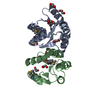


 Mass: 96.063 Da / Num. of mol.: 2 / Source method: obtained synthetically / Formula: SO4
 Mass: 62.068 Da / Num. of mol.: 6 / Source method: obtained synthetically / Formula: C2H6O2
 Mass: 18.015 Da / Num. of mol.: 367 / Source method: isolated from a natural source / Formula: H2O
 Sample preparation
 / Beamline: BL11-1 / Wavelength: 0.97916,0.91837,0.97876
 Processing