 Movie
Movie Controller


+ Open data

- Basic information
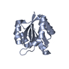
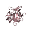
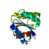
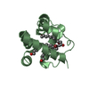
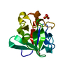
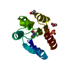
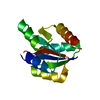
| Entry | Database: PDB / ID: 3grc | ||||||
|---|---|---|---|---|---|---|---|
| Title | Crystal structure of a sensor protein from Polaromonas sp. JS666 | ||||||
 Components Components | Sensor protein, Kinase | ||||||
 Keywords Keywords | TRANSFERASE / Sensor protein / Kinase / Protein Structure Initiative II(PSI II) / NYSGXRC / 11025b / Structural Genomics / New York SGX Research Center for Structural Genomics / Phosphoprotein | ||||||
| Function / homology |  Function and homology information Function and homology informationhistidine phosphotransfer kinase activity / phosphorelay sensor kinase activity / histidine kinase / ATP binding / plasma membrane Similarity search - Function | ||||||
| Biological species |  Polaromonas sp. JS666 (bacteria) Polaromonas sp. JS666 (bacteria) | ||||||
| Method |  X-RAY DIFFRACTION / SYNCHROTRON / SAD / Resolution: 2.21 Å X-RAY DIFFRACTION / SYNCHROTRON / SAD / Resolution: 2.21 Å | ||||||
 Authors Authors | Palani, K. / Kumaran, D. / Burley, S.K. / Swaminathan, S. / New York SGX Research Center for Structural Genomics (NYSGXRC) | ||||||
 Citation Citation | Journal: To be Published Title: Crystal structure of a sensor protein from Polaromonas sp. JS666 Authors: Palani, K. / Kumaran, D. / Burley, S.K. / Swaminathan, S. | ||||||
| History |
|
- Structure visualization
| Structure viewer | Molecule: MolmilJmol/JSmol |
|---|
- Downloads & links
-Download
| PDBx/mmCIF format | 3grc.cif.gz | 111.1 KB | Display | PDBx/mmCIF format |
|---|---|---|---|---|
| PDB format | pdb3grc.ent.gz | 87.6 KB | Display | PDB format |
| PDBx/mmJSON format | 3grc.json.gz | Tree view | PDBx/mmJSON format | |
| Others |  Other downloads Other downloads |
-Validation report
| Arichive directory | https://data.pdbj.org/pub/pdb/validation_reports/gr/3grcftp://data.pdbj.org/pub/pdb/validation_reports/gr/3grc | HTTPS FTP |
|---|
-Related structure data
| Similar structure data | |
|---|---|
| Other databases |


-Links
 PDBj
PDBj


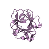
- Assembly
| Deposited unit | 
| ||||||||
|---|---|---|---|---|---|---|---|---|---|
| 1 | 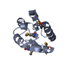
| ||||||||
| 2 | 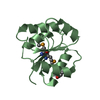
| ||||||||
| 3 | 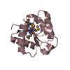
| ||||||||
| 4 | 
| ||||||||
| 5 |
| ||||||||
| Unit cell |
|
-Components
| #1: Protein | Mass: 15927.300 Da / Num. of mol.: 4 Source method: isolated from a genetically manipulated source Source: (gene. exp.) Polaromonas sp. JS666 (bacteria) / Gene: Bpro_4825 / Plasmid: BC-pSGX3 (BC) / Production host: #2: Water | ChemComp-HOH / |  Mass: 18.015 Da / Num. of mol.: 192 / Source method: isolated from a natural source / Formula: H2O Mass: 18.015 Da / Num. of mol.: 192 / Source method: isolated from a natural source / Formula: H2OHas protein modification | Y | |
|---|
-Experimental details
-Experiment
| Experiment | Method: X-RAY DIFFRACTION / Number of used crystals: 1 |
|---|
- Sample preparation
| Crystal | Density Matthews: 2.3 Å3/Da / Density % sol: 46.58 % |
|---|---|
| Crystal grow | Temperature: 298 K / Method: vapor diffusion, sitting drop / pH: 7 Details: 0.1M Sodium formate 12% Polyethylene glycol 3350, pH 7.0, VAPOR DIFFUSION, SITTING DROP, temperature 298.0K |
-Data collection
| Diffraction | Mean temperature: 100 K |
|---|---|
| Diffraction source | Source: SYNCHROTRON / Site: NSLS  / Beamline: X29A / Wavelength: 0.9792 Å / Beamline: X29A / Wavelength: 0.9792 Å |
| Detector | Type: ADSC QUANTUM 315 / Detector: CCD / Date: Mar 23, 2009 |
| Radiation | Monochromator: Si(III) CHANNEL / Protocol: SINGLE WAVELENGTH / Monochromatic (M) / Laue (L): M / Scattering type: x-ray |
| Radiation wavelength | Wavelength: 0.9792 Å / Relative weight: 1 |
| Reflection | Resolution: 2.21→40.44 Å / Num. all: 29767 / Num. obs: 29767 / % possible obs: 99 % / Observed criterion σ(F): 0 / Observed criterion σ(I): 0 / Redundancy: 12.8 % / Biso Wilson estimate: 24 Å2 / Rmerge(I) obs: 0.117 / Net I/σ(I): 13.6 |
| Reflection shell | Resolution: 2.2→2.28 Å / Redundancy: 10 % / Rmerge(I) obs: 0.393 / Mean I/σ(I) obs: 2 / Num. unique all: 2808 / % possible all: 96.2 |
- Processing
| Software |
| |||||||||||||||||||||||||
|---|---|---|---|---|---|---|---|---|---|---|---|---|---|---|---|---|---|---|---|---|---|---|---|---|---|---|
| Refinement | Method to determine structure: SAD / Resolution: 2.21→40.44 Å / Rfactor Rfree error: 0.007 / Data cutoff high absF: 130619.9 / Data cutoff low absF: 0 / Isotropic thermal model: RESTRAINED / Cross valid method: THROUGHOUT / σ(F): 0 / Stereochemistry target values: Engh & Huber
| |||||||||||||||||||||||||
| Solvent computation | Solvent model: FLAT MODEL / Bsol: 32.0498 Å2 / ksol: 0.339086 e/Å3 | |||||||||||||||||||||||||
| Displacement parameters | Biso mean: 36.8 Å2
| |||||||||||||||||||||||||
| Refine analyze |
| |||||||||||||||||||||||||
| Refinement step | Cycle: LAST / Resolution: 2.21→40.44 Å
| |||||||||||||||||||||||||
| Refine LS restraints |
| |||||||||||||||||||||||||
| LS refinement shell | Resolution: 2.2→2.34 Å / Rfactor Rfree error: 0.022 / Total num. of bins used: 6
| |||||||||||||||||||||||||
| Xplor file |
|
