 Movie
Movie Controller

[English] 日本語
 Yorodumi
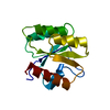
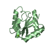
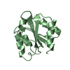
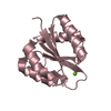
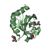
- PDB-4h60: High resolution structure of Vibrio cholerae chemotaxis protein C... -
+ Open data

- Basic information
| Entry | Database: PDB / ID: 4h60 | ||||||
|---|---|---|---|---|---|---|---|
| Title | High resolution structure of Vibrio cholerae chemotaxis protein CheY4 crystallized in low pH (4.0) condition | ||||||
 Components Components | Chemotaxis protein CheY | ||||||
 Keywords Keywords | SIGNALING PROTEIN / Rossmann Fold / response regulator / chemotaxis | ||||||
| Function / homology | Response regulator / Rossmann fold / 3-Layer(aba) Sandwich / Alpha Beta / :  Function and homology information Function and homology information | ||||||
| Biological species |   Vibrio cholerae (bacteria) Vibrio cholerae (bacteria) | ||||||
| Method |  X-RAY DIFFRACTION / MOLECULAR REPLACEMENT / Resolution: 1.66 Å X-RAY DIFFRACTION / MOLECULAR REPLACEMENT / Resolution: 1.66 Å | ||||||
 Authors Authors | Biswas, M. / Dasgupta, J. / Sen, U. | ||||||
 Citation Citation | Journal: Plos One / Year: 2013 Title: Conformational Barrier of CheY3 and Inability of CheY4 to Bind FliM Control the Flagellar Motor Action in Vibrio cholerae. Authors: Biswas, M. / Dey, S. / Khamrui, S. / Sen, U. / Dasgupta, J. #1: Journal: Acta Crystallogr.,Sect.F / Year: 2011 Title: Overexpression, purification, crystallization and preliminary X-ray analysis of CheY4 from Vibrio cholerae O395. Authors: Biswas, M. / Khamrui, S. / Sen, U. / Dasgupta, J. | ||||||
| History |
|
- Structure visualization
| Structure viewer | Molecule: MolmilJmol/JSmol |
|---|
- Downloads & links
-Download
| PDBx/mmCIF format | 4h60.cif.gz | 40.2 KB | Display | PDBx/mmCIF format |
|---|---|---|---|---|
| PDB format | pdb4h60.ent.gz | 26.1 KB | Display | PDB format |
| PDBx/mmJSON format | 4h60.json.gz | Tree view | PDBx/mmJSON format | |
| Others |  Other downloads Other downloads |
-Validation report
| Arichive directory | https://data.pdbj.org/pub/pdb/validation_reports/h6/4h60ftp://data.pdbj.org/pub/pdb/validation_reports/h6/4h60 | HTTPS FTP |
|---|
-Related structure data
| Related structure data | 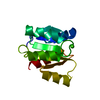 3to5SC 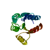 4hnqC 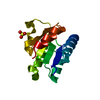 4hnrC 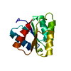 4hnsC 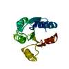 4jp1C 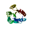 4lx8C S: Starting model for refinement C: citing same article ( |
|---|---|
| Similar structure data |


-Links
 PDBj
PDBj- Assembly
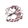
| Deposited unit | 
| ||||||||
|---|---|---|---|---|---|---|---|---|---|
| 1 |
| ||||||||
| Unit cell |
|
-Components
| #1: Protein | Mass: 13290.412 Da / Num. of mol.: 1 / Fragment: UNP residues 7-125 Source method: isolated from a genetically manipulated source Source: (gene. exp.) Vibrio cholerae (bacteria) / Strain: ATCC 39541 / Ogawa 395 / O395 / Gene: cheY-4 / Production host: |
|---|---|
| #2: Chemical | ChemComp-CA /   Mass: 40.078 Da / Num. of mol.: 1 / Source method: obtained synthetically / Formula: Ca Mass: 40.078 Da / Num. of mol.: 1 / Source method: obtained synthetically / Formula: Ca |
| #3: Chemical | ChemComp-SO4 /   Mass: 96.063 Da / Num. of mol.: 1 / Source method: obtained synthetically / Formula: SO4 Mass: 96.063 Da / Num. of mol.: 1 / Source method: obtained synthetically / Formula: SO4 |
| #4: Water | ChemComp-HOH /  Mass: 18.015 Da / Num. of mol.: 113 / Source method: isolated from a natural source / Formula: H2O Mass: 18.015 Da / Num. of mol.: 113 / Source method: isolated from a natural source / Formula: H2O |
-Experimental details
-Experiment
| Experiment | Method: X-RAY DIFFRACTION / Number of used crystals: 1 |
|---|
- Sample preparation
| Crystal | Density Matthews: 1.81 Å3/Da / Density % sol: 31.96 % |
|---|---|
| Crystal grow | Temperature: 293 K / Method: vapor diffusion, hanging drop / pH: 4 Details: 1.6 M ammonium sulfate, pH 4.0, VAPOR DIFFUSION, HANGING DROP, temperature 293K |
-Data collection
| Diffraction | Mean temperature: 100 K |
|---|---|
| Diffraction source | Source: ROTATING ANODE / Type: RIGAKU RU300 / Wavelength: 1.54 Å |
| Detector | Type: MAR scanner 345 mm plate / Detector: IMAGE PLATE / Date: Jan 1, 2004 |
| Radiation | Monochromator: GRAPHITE / Protocol: SINGLE WAVELENGTH / Monochromatic (M) / Laue (L): M / Scattering type: x-ray |
| Radiation wavelength | Wavelength: 1.54 Å / Relative weight: 1 |
| Reflection | Resolution: 1.66→30 Å / Num. all: 11447 / Num. obs: 10882 / % possible obs: 95.1 % / Observed criterion σ(F): 0 / Observed criterion σ(I): 0 / Biso Wilson estimate: 18.4 Å2 / Rmerge(I) obs: 0.0449 / Net I/σ(I): 8.6 |
| Reflection shell | Highest resolution: 1.66 Å |
- Processing
| Software |
| ||||||||||||||||||||||||||||||||||||
|---|---|---|---|---|---|---|---|---|---|---|---|---|---|---|---|---|---|---|---|---|---|---|---|---|---|---|---|---|---|---|---|---|---|---|---|---|---|
| Refinement | Method to determine structure: MOLECULAR REPLACEMENT Starting model: PDB ENTRY 3TO5 Resolution: 1.66→22.24 Å / Rfactor Rfree error: 0.01 / Data cutoff high absF: 861015.34 / Data cutoff low absF: 0 / Isotropic thermal model: RESTRAINED / Cross valid method: THROUGHOUT / σ(F): 0 / Stereochemistry target values: Engh & Huber / Details: BULK SOLVENT MODEL USED
| ||||||||||||||||||||||||||||||||||||
| Solvent computation | Solvent model: FLAT MODEL / Bsol: 44.7319 Å2 / ksol: 0.4 e/Å3 | ||||||||||||||||||||||||||||||||||||
| Displacement parameters | Biso mean: 20.3 Å2
| ||||||||||||||||||||||||||||||||||||
| Refine analyze |
| ||||||||||||||||||||||||||||||||||||
| Refinement step | Cycle: LAST / Resolution: 1.66→22.24 Å
| ||||||||||||||||||||||||||||||||||||
| Refine LS restraints |
| ||||||||||||||||||||||||||||||||||||
| LS refinement shell | Resolution: 1.66→1.76 Å / Rfactor Rfree error: 0.032 / Total num. of bins used: 6
| ||||||||||||||||||||||||||||||||||||
| Xplor file |
|
