 Movie
Movie Controller

[English] 日本語
 Yorodumi
- PDB-2eio: Design of Disulfide-linked Thioredoxin Dimers and Multimers Throu... -
+ Open data

- Basic information
| Entry | Database: PDB / ID: 2eio | ||||||
|---|---|---|---|---|---|---|---|
| Title | Design of Disulfide-linked Thioredoxin Dimers and Multimers Through Analysis of Crystal Contacts | ||||||
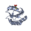
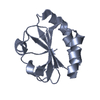
 Components Components | Thioredoxin 1 | ||||||
 Keywords Keywords | ELECTRON TRANSPORT / THIOREDOXIN / MUTANT / DI-SULFIDE BOND | ||||||
| Function / homology |  Function and homology information Function and homology informationDNA polymerase processivity factor activity / protein-disulfide reductase activity / cell redox homeostasis / cytoplasm / cytosol Similarity search - Function | ||||||
| Biological species |  | ||||||
| Method |  X-RAY DIFFRACTION / SYNCHROTRON / MOLECULAR REPLACEMENT / Resolution: 2.6 Å X-RAY DIFFRACTION / SYNCHROTRON / MOLECULAR REPLACEMENT / Resolution: 2.6 Å | ||||||
 Authors Authors | Kobayashi, M. | ||||||
 Citation Citation | Journal: J.Mol.Biol. / Year: 2007 Title: Design of Disulfide-linked Thioredoxin Dimers and Multimers Through Analysis of Crystal Contacts Authors: Das, M. / Kobayashi, M. / Yamada, Y. / Sreeramulu, S. / Ramakrishnan, C. / Wakatsuki, S. / Kato, R. / Varadarajan, R. | ||||||
| History |
|
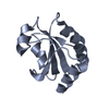
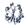
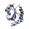
- Structure visualization
| Structure viewer | Molecule: MolmilJmol/JSmol |
|---|
- Downloads & links
-Download
| PDBx/mmCIF format | 2eio.cif.gz | 91.2 KB | Display | PDBx/mmCIF format |
|---|---|---|---|---|
| PDB format | pdb2eio.ent.gz | 70.3 KB | Display | PDB format |
| PDBx/mmJSON format | 2eio.json.gz | Tree view | PDBx/mmJSON format | |
| Others |  Other downloads Other downloads |
-Validation report
| Arichive directory | https://data.pdbj.org/pub/pdb/validation_reports/ei/2eioftp://data.pdbj.org/pub/pdb/validation_reports/ei/2eio | HTTPS FTP |
|---|
-Related structure data
| Related structure data |  2eiqC  2eirC 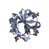 2trxS C: citing same article ( S: Starting model for refinement |
|---|---|
| Similar structure data |


-Links
 PDBj
PDBj

- Assembly
| Deposited unit | 
| ||||||||
|---|---|---|---|---|---|---|---|---|---|
| 1 | 
| ||||||||
| 2 | 
| ||||||||
| 3 | 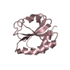
| ||||||||
| 4 | 
| ||||||||
| Unit cell |
|
-Components
| #1: Protein | Mass: 11661.417 Da / Num. of mol.: 4 / Mutation: E101C Source method: isolated from a genetically manipulated source Source: (gene. exp.) #2: Water | ChemComp-HOH / |  Mass: 18.015 Da / Num. of mol.: 57 / Source method: isolated from a natural source / Formula: H2O Mass: 18.015 Da / Num. of mol.: 57 / Source method: isolated from a natural source / Formula: H2OHas protein modification | Y | |
|---|
-Experimental details
-Experiment
| Experiment | Method: X-RAY DIFFRACTION / Number of used crystals: 1 |
|---|
- Sample preparation
| Crystal | Density Matthews: 2.42 Å3/Da / Density % sol: 49.22 % |
|---|---|
| Crystal grow | Temperature: 289 K / Method: vapor diffusion, hanging drop / pH: 8.5 Details: 20% PEG 3350, 0.1M TRIS, 0.2M MAGNESIUM CHLORIDE, pH 8.50, VAPOR DIFFUSION, HANGING DROP, temperature 289K |
-Data collection
| Diffraction | Mean temperature: 100 K |
|---|---|
| Diffraction source | Source: SYNCHROTRON / Site: Photon Factory  / Beamline: AR-NW12A / Wavelength: 1 / Beamline: AR-NW12A / Wavelength: 1 |
| Detector | Type: ADSC QUANTUM 210 / Detector: CCD / Date: Feb 17, 2005 / Details: MIRROR |
| Radiation | Monochromator: Si(111) / Protocol: SINGLE WAVELENGTH / Monochromatic (M) / Laue (L): M / Scattering type: x-ray |
| Radiation wavelength | Wavelength: 1 Å / Relative weight: 1 |
| Reflection | Resolution: 2.6→50 Å / Num. obs: 14472 / % possible obs: 100 % / Redundancy: 7.2 % / Biso Wilson estimate: 27.7 Å2 / Rmerge(I) obs: 0.081 / Rsym value: 0.081 / Net I/σ(I): 24.3 |
| Reflection shell | Resolution: 2.6→2.69 Å / Redundancy: 7.3 % / Rmerge(I) obs: 0.258 / Mean I/σ(I) obs: 6.7 / Rsym value: 0.258 / % possible all: 99.9 |
- Processing
| Software |
| ||||||||||||||||||||||||||||||||||||||||||||||||||||||||||||||||||||||||||||||||
|---|---|---|---|---|---|---|---|---|---|---|---|---|---|---|---|---|---|---|---|---|---|---|---|---|---|---|---|---|---|---|---|---|---|---|---|---|---|---|---|---|---|---|---|---|---|---|---|---|---|---|---|---|---|---|---|---|---|---|---|---|---|---|---|---|---|---|---|---|---|---|---|---|---|---|---|---|---|---|---|---|---|
| Refinement | Method to determine structure: MOLECULAR REPLACEMENT Starting model: PDB ENTRY 2TRX Resolution: 2.6→38.58 Å / Rfactor Rfree error: 0.008 / Data cutoff high absF: 148038.94 / Data cutoff low absF: 0 / Cross valid method: THROUGHOUT / σ(F): 0
| ||||||||||||||||||||||||||||||||||||||||||||||||||||||||||||||||||||||||||||||||
| Solvent computation | Solvent model: FLAT MODEL / Bsol: 21.8641 Å2 / ksol: 0.35 e/Å3 | ||||||||||||||||||||||||||||||||||||||||||||||||||||||||||||||||||||||||||||||||
| Displacement parameters | Biso mean: 24 Å2
| ||||||||||||||||||||||||||||||||||||||||||||||||||||||||||||||||||||||||||||||||
| Refine analyze |
| ||||||||||||||||||||||||||||||||||||||||||||||||||||||||||||||||||||||||||||||||
| Refinement step | Cycle: LAST / Resolution: 2.6→38.58 Å
| ||||||||||||||||||||||||||||||||||||||||||||||||||||||||||||||||||||||||||||||||
| Refine LS restraints |
| ||||||||||||||||||||||||||||||||||||||||||||||||||||||||||||||||||||||||||||||||
| LS refinement shell | Resolution: 2.6→2.76 Å / Rfactor Rfree error: 0.024 / Total num. of bins used: 6
| ||||||||||||||||||||||||||||||||||||||||||||||||||||||||||||||||||||||||||||||||
| Xplor file |
|
