 Movie
Movie Controller

[English] 日本語
 Yorodumi
- PDB-2h9b: Crystal structure of the effector binding domain of a BenM varian... -
+ Open data

- Basic information
| Entry | Database: PDB / ID: 2h9b | ||||||
|---|---|---|---|---|---|---|---|
| Title | Crystal structure of the effector binding domain of a BenM variant (BenM R156H/T157S) | ||||||
 Components Components | HTH-type transcriptional regulator benM | ||||||
 Keywords Keywords | TRANSCRIPTION / LTTR / BenM / Transcriptional activator / lysR-type transcriptional regulator | ||||||
| Function / homology |  Function and homology information Function and homology informationprotein-DNA complex / DNA-binding transcription factor activity / DNA-templated transcription / DNA binding Similarity search - Function | ||||||
| Biological species |  Acinetobacter sp. (bacteria) Acinetobacter sp. (bacteria) | ||||||
| Method |  X-RAY DIFFRACTION / SYNCHROTRON / MOLECULAR REPLACEMENT / Resolution: 1.8 Å X-RAY DIFFRACTION / SYNCHROTRON / MOLECULAR REPLACEMENT / Resolution: 1.8 Å | ||||||
 Authors Authors | Ezezika, O.C. / Craven, S.H. / Neidle, E.L. / Momany, C. | ||||||
 Citation Citation | Journal: Mol.Microbiol. / Year: 2009 Title: Inducer responses of BenM, a LysR-type transcriptional regulator from Acinetobacter baylyi ADP1. Authors: Craven, S.H. / Ezezika, O.C. / Haddad, S. / Hall, R.A. / Momany, C. / Neidle, E.L. #1: Journal: To be PublishedTitle: Distinct Effector-binding Sites Enable Synergistic Transcriptional Activation By BenM, a LysR-type Regulator Authors: Ezezika, O.C. / Haddad, S. / Clark, T.J. / Neidle, E.L. / Momany, C. | ||||||
| History |
|
- Structure visualization
| Structure viewer | Molecule: MolmilJmol/JSmol |
|---|
- Downloads & links
-Download
| PDBx/mmCIF format | 2h9b.cif.gz | 122.5 KB | Display | PDBx/mmCIF format |
|---|---|---|---|---|
| PDB format | pdb2h9b.ent.gz | 91.2 KB | Display | PDB format |
| PDBx/mmJSON format | 2h9b.json.gz | Tree view | PDBx/mmJSON format | |
| Others |  Other downloads Other downloads |
-Validation report
| Arichive directory | https://data.pdbj.org/pub/pdb/validation_reports/h9/2h9bftp://data.pdbj.org/pub/pdb/validation_reports/h9/2h9b | HTTPS FTP |
|---|
-Related structure data
| Related structure data |  2h99C  3glbC  2f97S S: Starting model for refinement C: citing same article ( |
|---|---|
| Similar structure data |


-Links
 PDBj
PDBj- Assembly
| Deposited unit | 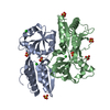
| ||||||||
|---|---|---|---|---|---|---|---|---|---|
| 1 |
| ||||||||
| Unit cell |
| ||||||||
| Details | biological unit is a dimer |
-Components
| #1: Protein | Mass: 35611.961 Da / Num. of mol.: 2 / Fragment: Effector binding domain / Mutation: R156H, T157S Source method: isolated from a genetically manipulated source Source: (gene. exp.) Acinetobacter sp. (bacteria) / Strain: ADP1 / Gene: benM, benR / Plasmid: pET21B / Species (production host): Escherichia coli / Production host: #2: Chemical | ChemComp-SO4 /   Mass: 96.063 Da / Num. of mol.: 7 / Source method: obtained synthetically / Formula: SO4 Mass: 96.063 Da / Num. of mol.: 7 / Source method: obtained synthetically / Formula: SO4#3: Chemical |   Mass: 35.453 Da / Num. of mol.: 3 / Source method: obtained synthetically / Formula: Cl Mass: 35.453 Da / Num. of mol.: 3 / Source method: obtained synthetically / Formula: Cl#4: Water | ChemComp-HOH / |  Mass: 18.015 Da / Num. of mol.: 741 / Source method: isolated from a natural source / Formula: H2O Mass: 18.015 Da / Num. of mol.: 741 / Source method: isolated from a natural source / Formula: H2O |
|---|
-Experimental details
-Experiment
| Experiment | Method: X-RAY DIFFRACTION / Number of used crystals: 1 |
|---|
- Sample preparation
| Crystal | Density Matthews: 2.5 Å3/Da / Density % sol: 49.9 % |
|---|---|
| Crystal grow | Temperature: 296.4 K Details: Precipitant:2.0 M ammonium sulfate Protein: 20 mM tris HCl, 0.5 M NaCl, pH 7.9, 10% glycerol Equal volumes mixed, Microbatch under oil, temperature 296.4K |
-Data collection
| Diffraction | Mean temperature: 100 K |
|---|---|
| Diffraction source | Source: SYNCHROTRON / Site: APS  / Beamline: 22-BM / Wavelength: 1 Å / Beamline: 22-BM / Wavelength: 1 Å |
| Detector | Type: MARRESEARCH / Detector: CCD / Date: Aug 22, 2005 |
| Radiation | Protocol: SINGLE WAVELENGTH / Monochromatic (M) / Laue (L): M / Scattering type: x-ray |
| Radiation wavelength | Wavelength: 1 Å / Relative weight: 1 |
| Reflection | Resolution: 1.8→50 Å / Num. obs: 48633 / % possible obs: 100 % / Redundancy: 6.9 % / Rmerge(I) obs: 0.078 / Χ2: 0.858 |
| Reflection shell | Resolution: 1.8→1.86 Å / % possible obs: 99.9 % / Redundancy: 6.3 % / Rmerge(I) obs: 0.464 / Num. unique obs: 4763 / Χ2: 0.799 / % possible all: 99.77 |
- Processing
| Software |
| |||||||||||||||||||||||||||||||||||||||||||||||||||||||||||||||||||||||||||||||||||||||||||||||||||||||||||||||||||||||||||||
|---|---|---|---|---|---|---|---|---|---|---|---|---|---|---|---|---|---|---|---|---|---|---|---|---|---|---|---|---|---|---|---|---|---|---|---|---|---|---|---|---|---|---|---|---|---|---|---|---|---|---|---|---|---|---|---|---|---|---|---|---|---|---|---|---|---|---|---|---|---|---|---|---|---|---|---|---|---|---|---|---|---|---|---|---|---|---|---|---|---|---|---|---|---|---|---|---|---|---|---|---|---|---|---|---|---|---|---|---|---|---|---|---|---|---|---|---|---|---|---|---|---|---|---|---|---|---|
| Refinement | Method to determine structure: MOLECULAR REPLACEMENT Starting model: PDB accession code 2F97, BenM-EBD (high pH) Resolution: 1.8→46.7 Å / Cor.coef. Fo:Fc: 0.957 / Cor.coef. Fo:Fc free: 0.935 / SU B: 4.306 / SU ML: 0.074 / Cross valid method: THROUGHOUT / σ(F): 0 / σ(I): 0 / ESU R: 0.119 / ESU R Free: 0.117 / Stereochemistry target values: Engh & Huber / Details: HYDROGENS HAVE BEEN ADDED IN THE RIDING POSITIONS
| |||||||||||||||||||||||||||||||||||||||||||||||||||||||||||||||||||||||||||||||||||||||||||||||||||||||||||||||||||||||||||||
| Solvent computation | Ion probe radii: 0.8 Å / Shrinkage radii: 0.8 Å / VDW probe radii: 1.2 Å / Solvent model: BABINET MODEL WITH MASK | |||||||||||||||||||||||||||||||||||||||||||||||||||||||||||||||||||||||||||||||||||||||||||||||||||||||||||||||||||||||||||||
| Displacement parameters | Biso mean: 16.201 Å2
| |||||||||||||||||||||||||||||||||||||||||||||||||||||||||||||||||||||||||||||||||||||||||||||||||||||||||||||||||||||||||||||
| Refinement step | Cycle: LAST / Resolution: 1.8→46.7 Å
| |||||||||||||||||||||||||||||||||||||||||||||||||||||||||||||||||||||||||||||||||||||||||||||||||||||||||||||||||||||||||||||
| Refine LS restraints |
| |||||||||||||||||||||||||||||||||||||||||||||||||||||||||||||||||||||||||||||||||||||||||||||||||||||||||||||||||||||||||||||
| LS refinement shell | Resolution: 1.8→1.846 Å / Total num. of bins used: 20
|
