 Movie
Movie Controller

[English] 日本語
 Yorodumi
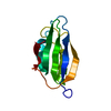
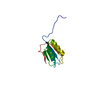
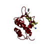
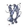
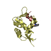
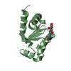
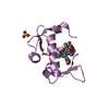
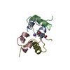
- PDB-2gv1: NMR solution structure of the Acylphosphatase from Eschaerichia Coli -
+ Open data

- Basic information
| Entry | Database: PDB / ID: 2gv1 | ||||||
|---|---|---|---|---|---|---|---|
| Title | NMR solution structure of the Acylphosphatase from Eschaerichia Coli | ||||||
 Components Components | Probable acylphosphatase | ||||||
 Keywords Keywords | HYDROLASE / globular alpha-helix/beta-sheet protein | ||||||
| Function / homology |  Function and homology information Function and homology information | ||||||
| Biological species |  | ||||||
| Method | SOLUTION NMR / torsion angle dynamics | ||||||
 Authors Authors | Pagano, K. / Corazza, A. / Viglino, P. / Esposito, G. | ||||||
 Citation Citation | Journal: J.Biomol.Nmr / Year: 2006 Title: NMR solution structure of the acylphosphatase from Escherichia coli. Authors: Pagano, K. / Ramazzotti, M. / Viglino, P. / Esposito, G. / Degl'innocenti, D. / Taddei, N. / Corazza, A. #1: Journal: Proteins / Year: 2006Title: Structure, Conformational Stability, and Enzymatic Properties of Acylphosphatase from the Hyperthermophile Sulfolobus Solfataricus Authors: Corazza, A. / Rosano, C. / Pagano, K. / Alverdi, V. / Esposito, G. / Capanni, C. / Bemporad, F. / Plakoutsi, G. / Stefani, M. / Chiti, F. / Zuccotti, S. / Bolognesi, M. / Viglino, P. #2: Journal: Structure / Year: 1997Title: Crystal structure of common type acylphosphatase from bovine testis Authors: Thunnissen, M.M. / Taddei, N. / Liguri, G. / Ramponi, G. / Nordlund, P. #3: Journal: J.Mol.Biol. / Year: 1992Title: Three-dimensional structure of acylphosphatase. Refinement and structure analysis. Authors: Pastore, A. / Saudek, V. / Ramponi, G. / Williams, R.J. | ||||||
| History |
|
- Structure visualization
| Structure viewer | Molecule: MolmilJmol/JSmol |
|---|
- Downloads & links
-Download
| PDBx/mmCIF format | 2gv1.cif.gz | 561.9 KB | Display | PDBx/mmCIF format |
|---|---|---|---|---|
| PDB format | pdb2gv1.ent.gz | 471.9 KB | Display | PDB format |
| PDBx/mmJSON format | 2gv1.json.gz | Tree view | PDBx/mmJSON format | |
| Others |  Other downloads Other downloads |
-Validation report
| Arichive directory | https://data.pdbj.org/pub/pdb/validation_reports/gv/2gv1ftp://data.pdbj.org/pub/pdb/validation_reports/gv/2gv1 | HTTPS FTP |
|---|
-Related structure data
| Related structure data | |
|---|---|
| Similar structure data |


-Links
 PDBj
PDBj- Assembly
| Deposited unit | 
| |||||||||
|---|---|---|---|---|---|---|---|---|---|---|
| 1 |
| |||||||||
| NMR ensembles |
|
-Components
| #1: Protein | Mass: 10314.760 Da / Num. of mol.: 1 Source method: isolated from a genetically manipulated source Source: (gene. exp.) |
|---|---|
| Has protein modification | Y |
-Experimental details
-Experiment
| Experiment | Method: SOLUTION NMR | ||||||||||||||||||||||||
|---|---|---|---|---|---|---|---|---|---|---|---|---|---|---|---|---|---|---|---|---|---|---|---|---|---|
| NMR experiment |
| ||||||||||||||||||||||||
| NMR details | Text: The structure was determined using triple-resonance NMR spectroscopy and standard 2D homonuclear techniques |
 HSQC
HSQC- Sample preparation
| Details |
| |||||||||
|---|---|---|---|---|---|---|---|---|---|---|
| Sample conditions | Ionic strength: 65 mM / pH: 4.95 / Pressure: ambient / Temperature: 310 K |
-NMR measurement
| Radiation | Protocol: SINGLE WAVELENGTH / Monochromatic (M) / Laue (L): M |
|---|---|
| Radiation wavelength | Relative weight: 1 |
| NMR spectrometer | Type: Bruker AVANCE / Manufacturer: Bruker / Model: AVANCE / Field strength: 500 MHz |
- Processing
| NMR software |
| ||||||||||||||||||||
|---|---|---|---|---|---|---|---|---|---|---|---|---|---|---|---|---|---|---|---|---|---|
| Refinement | Method: torsion angle dynamics / Software ordinal: 1 Details: the structures are based on a total of 1029 restraints, 970 are NOE-derived distance restraints and 59 dihedral angle restraints | ||||||||||||||||||||
| NMR representative | Selection criteria: fewest violations | ||||||||||||||||||||
| NMR ensemble | Conformer selection criteria: target function / Conformers calculated total number: 380 / Conformers submitted total number: 20 |
