 Movie
Movie Controller

[English] 日本語
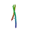
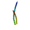
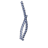
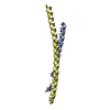
 Yorodumi
- PDB-2gl2: Crystal structure of the tetra mutant (T66G,R67G,F68G,Y69G) of ba... -
+ Open data

- Basic information
| Entry | Database: PDB / ID: 2gl2 | ||||||
|---|---|---|---|---|---|---|---|
| Title | Crystal structure of the tetra mutant (T66G,R67G,F68G,Y69G) of bacterial adhesin FadA | ||||||
 Components Components | adhesion A | ||||||
 Keywords Keywords | CELL ADHESION / antiparallel helix-loop-helix / FadA Gly4 mutant | ||||||
| Function / homology | Adhesion protein FadA / Adhesion protein FadA / Helix Hairpins - #1700 / : / Helix Hairpins / Orthogonal Bundle / Mainly Alpha / : / Adhesion A Function and homology information Function and homology information | ||||||
| Biological species |  Fusobacterium nucleatum (bacteria) Fusobacterium nucleatum (bacteria) | ||||||
| Method |  X-RAY DIFFRACTION / SYNCHROTRON / MOLECULAR REPLACEMENT / Resolution: 2.5 Å X-RAY DIFFRACTION / SYNCHROTRON / MOLECULAR REPLACEMENT / Resolution: 2.5 Å | ||||||
 Authors Authors | Nithianantham, S. / Xu, M. / Wu, N. / Shoham, M. / Han, Y.W. | ||||||
 Citation Citation | Journal: Acta Crystallogr.,Sect.F / Year: 2006 Title: Crystallization and preliminary X-ray data of the FadA adhesin from Fusobacterium nucleatum. Authors: Nithianantham, S. / Xu, M. / Wu, N. / Han, Y.W. / Shoham, M. | ||||||
| History |
|
- Structure visualization
| Structure viewer | Molecule: MolmilJmol/JSmol |
|---|
- Downloads & links
-Download
| PDBx/mmCIF format | 2gl2.cif.gz | 58.3 KB | Display | PDBx/mmCIF format |
|---|---|---|---|---|
| PDB format | pdb2gl2.ent.gz | 42.8 KB | Display | PDB format |
| PDBx/mmJSON format | 2gl2.json.gz | Tree view | PDBx/mmJSON format | |
| Others |  Other downloads Other downloads |
-Validation report
| Summary document | 2gl2_validation.pdf.gz | 429 KB | Display | wwPDB validaton report |
|---|---|---|---|---|
| Full document | 2gl2_full_validation.pdf.gz | 432.7 KB | Display | |
| Data in XML | 2gl2_validation.xml.gz | 12.6 KB | Display | |
| Data in CIF | 2gl2_validation.cif.gz | 17.8 KB | Display | |
| Arichive directory | https://data.pdbj.org/pub/pdb/validation_reports/gl/2gl2ftp://data.pdbj.org/pub/pdb/validation_reports/gl/2gl2 | HTTPS FTP |
-Related structure data
| Related structure data |  2avr S: Starting model for refinement |
|---|---|
| Similar structure data |

-Links
 PDBj
PDBj- Assembly
| Deposited unit | 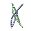
| ||||||||
|---|---|---|---|---|---|---|---|---|---|
| 1 | 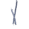
| ||||||||
| 2 | 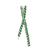
| ||||||||
| Unit cell |
|
-Components
| #1: Protein | Mass: 13329.401 Da / Num. of mol.: 2 / Fragment: residues 19-129 / Mutation: T66G, R67G, F68G, Y69G Source method: isolated from a genetically manipulated source Source: (gene. exp.) Fusobacterium nucleatum (bacteria) / Plasmid: pYH1378 / Species (production host): Escherichia coli / Production host: #2: Water | ChemComp-HOH / |  Mass: 18.015 Da / Num. of mol.: 223 / Source method: isolated from a natural source / Formula: H2O Mass: 18.015 Da / Num. of mol.: 223 / Source method: isolated from a natural source / Formula: H2O |
|---|
-Experimental details
-Experiment
| Experiment | Method: X-RAY DIFFRACTION / Number of used crystals: 1 |
|---|
- Sample preparation
| Crystal | Density Matthews: 3.57 Å3/Da / Density % sol: 65.53 % |
|---|---|
| Crystal grow | Temperature: 295 K / Method: vapor diffusion / pH: 4.6 Details: Sodium acetate, beta-octyl glucoside, pH 4.6, VAPOR DIFFUSION, temperature 295K |
-Data collection
| Diffraction | Mean temperature: 100 K |
|---|---|
| Diffraction source | Source: SYNCHROTRON / Site: APS  / Beamline: 19-BM / Wavelength: 0.97899 Å / Beamline: 19-BM / Wavelength: 0.97899 Å |
| Detector | Type: CUSTOM-MADE / Detector: CCD / Date: Mar 1, 2006 / Details: Mirrors |
| Radiation | Monochromator: GRAPHITE / Protocol: SINGLE WAVELENGTH / Monochromatic (M) / Laue (L): M / Scattering type: x-ray |
| Radiation wavelength | Wavelength: 0.97899 Å / Relative weight: 1 |
| Reflection | Resolution: 2.5→50 Å / Num. all: 11518 / Num. obs: 11518 / % possible obs: 89.4 % / Observed criterion σ(F): 0 / Observed criterion σ(I): 0 / Redundancy: 4.7 % / Rmerge(I) obs: 0.062 / Χ2: 1.243 / Net I/σ(I): 16.6 |
| Reflection shell | Resolution: 2.5→2.59 Å / % possible obs: 49.1 % / Redundancy: 1.8 % / Rmerge(I) obs: 0.56 / Num. unique obs: 632 / Χ2: 1.52 / % possible all: 48.5 |
- Processing
| Software |
| ||||||||||||||||||||||||||||
|---|---|---|---|---|---|---|---|---|---|---|---|---|---|---|---|---|---|---|---|---|---|---|---|---|---|---|---|---|---|
| Refinement | Method to determine structure: MOLECULAR REPLACEMENT Starting model: pdb entry 2AVR 2avr Resolution: 2.5→29.2 Å / Cross valid method: THROUGHOUT / σ(F): 0 / Stereochemistry target values: Engh & Huber
| ||||||||||||||||||||||||||||
| Solvent computation | Bsol: 34.271 Å2 | ||||||||||||||||||||||||||||
| Displacement parameters | Biso mean: 46.112 Å2
| ||||||||||||||||||||||||||||
| Refine analyze | Luzzati coordinate error obs: 0.371 Å / Luzzati sigma a obs: 0.214 Å | ||||||||||||||||||||||||||||
| Refinement step | Cycle: LAST / Resolution: 2.5→29.2 Å
| ||||||||||||||||||||||||||||
| Refine LS restraints |
| ||||||||||||||||||||||||||||
| LS refinement shell | Resolution: 2.5→2.54 Å /
| ||||||||||||||||||||||||||||
| Xplor file |
|
