 Movie
Movie Controller


+ Open data

- Basic information
| Entry | Database: PDB / ID: 3etw | |||||||||
|---|---|---|---|---|---|---|---|---|---|---|
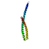
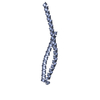
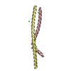
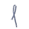
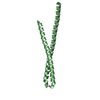
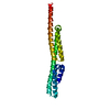
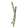
| Title | Crystal Structure of bacterial adhesin FadA | |||||||||
 Components Components | Adhesin A | |||||||||
 Keywords Keywords | CELL ADHESION / antiparallel helix-loop-helix / Leucine chain / cell adhesin | |||||||||
| Function / homology | Adhesion protein FadA / Adhesion protein FadA / Helix Hairpins - #1700 / : / Helix Hairpins / Orthogonal Bundle / Mainly Alpha / THIOCYANATE ION / Adhesion A Function and homology information Function and homology information | |||||||||
| Biological species |  Fusobacterium nucleatum (bacteria) Fusobacterium nucleatum (bacteria) | |||||||||
| Method |  X-RAY DIFFRACTION / SYNCHROTRON / MAD / Resolution: 2 Å X-RAY DIFFRACTION / SYNCHROTRON / MAD / Resolution: 2 Å | |||||||||
 Authors Authors | Nithianantham, S. / Xu, M. / Wu, N. / Shoham, M. / Han, Y.W. | |||||||||
 Citation Citation | Journal: J.Biol.Chem. / Year: 2009 Title: Crystal Structure of FadA Adhesin from Fusobacterium nucleatum Reveals a Novel Oligomerization Motif, the Leucine Chain. Authors: Nithianantham, S. / Xu, M. / Yamada, M. / Ikegami, A. / Shoham, M. / Han, Y.W. #1: Journal: Acta Crystallogr.,Sect.F / Year: 2006Title: Crystallization and preliminary X-ray data of the FadA adhesin from Fusobacterium nucleatum Authors: Nithianantham, S. / Xu, M. / Wu, N. / Han, Y.W. / Shoham, M. | |||||||||
| History |
|
- Structure visualization
| Structure viewer | Molecule: MolmilJmol/JSmol |
|---|
- Downloads & links
-Download
| PDBx/mmCIF format | 3etw.cif.gz | 38 KB | Display | PDBx/mmCIF format |
|---|---|---|---|---|
| PDB format | pdb3etw.ent.gz | 25.9 KB | Display | PDB format |
| PDBx/mmJSON format | 3etw.json.gz | Tree view | PDBx/mmJSON format | |
| Others |  Other downloads Other downloads |
-Validation report
| Arichive directory | https://data.pdbj.org/pub/pdb/validation_reports/et/3etwftp://data.pdbj.org/pub/pdb/validation_reports/et/3etw | HTTPS FTP |
|---|
-Related structure data


-Links
 PDBj
PDBj
- Assembly
| Deposited unit | 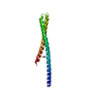
| ||||||||
|---|---|---|---|---|---|---|---|---|---|
| 1 |
| ||||||||
| Unit cell |
|
-Components
| #1: Protein | Mass: 13669.839 Da / Num. of mol.: 1 Source method: isolated from a genetically manipulated source Source: (gene. exp.) Fusobacterium nucleatum (bacteria) / Gene: fadA / Plasmid: pET21(b) / Production host: |
|---|---|
| #2: Chemical | ChemComp-SCN /   Mass: 58.082 Da / Num. of mol.: 1 / Source method: obtained synthetically / Formula: CNS Mass: 58.082 Da / Num. of mol.: 1 / Source method: obtained synthetically / Formula: CNS |
| #3: Water | ChemComp-HOH /  Mass: 18.015 Da / Num. of mol.: 162 / Source method: isolated from a natural source / Formula: H2O Mass: 18.015 Da / Num. of mol.: 162 / Source method: isolated from a natural source / Formula: H2O |
-Experimental details
-Experiment
| Experiment | Method: X-RAY DIFFRACTION / Number of used crystals: 1 |
|---|
- Sample preparation
| Crystal | Density Matthews: 4.7 Å3/Da / Density % sol: 73.82 % |
|---|---|
| Crystal grow | Temperature: 295 K / Method: vapor diffusion, sitting drop / pH: 5.6 Details: 0.1M Sodium citrate pH 5.6, 0.5M potassium thiocyanate, 5% dioxane, VAPOR DIFFUSION, SITTING DROP, temperature 295K |
-Data collection
| Diffraction |
| ||||||||||||||||||
|---|---|---|---|---|---|---|---|---|---|---|---|---|---|---|---|---|---|---|---|
| Diffraction source |
| ||||||||||||||||||
| Detector |
| ||||||||||||||||||
| Radiation |
| ||||||||||||||||||
| Radiation wavelength |
| ||||||||||||||||||
| Reflection | Resolution: 2→29.7 Å / Num. all: 17075 / Num. obs: 15871 / % possible obs: 93 % / Observed criterion σ(F): 0 / Observed criterion σ(I): 0 / Redundancy: 6.6 % / Biso Wilson estimate: 36.8 Å2 / Rmerge(I) obs: 0.045 / Χ2: 0.677 | ||||||||||||||||||
| Reflection shell | Resolution: 2→2.07 Å / Redundancy: 4.9 % / Rmerge(I) obs: 0.275 / Mean I/σ(I) obs: 2.813 / Num. unique all: 1068 / Χ2: 0.475 / % possible all: 63 |

-Phasing
| Phasing | Method: MAD | |||||||||||||||||||||||||||||||||||
|---|---|---|---|---|---|---|---|---|---|---|---|---|---|---|---|---|---|---|---|---|---|---|---|---|---|---|---|---|---|---|---|---|---|---|---|---|
| Phasing set |
| |||||||||||||||||||||||||||||||||||
| Phasing MAD set |
| |||||||||||||||||||||||||||||||||||
| Phasing MAD set site |
|
- Processing
| Software |
| ||||||||||||||||||||||||||||||||
|---|---|---|---|---|---|---|---|---|---|---|---|---|---|---|---|---|---|---|---|---|---|---|---|---|---|---|---|---|---|---|---|---|---|
| Refinement | Method to determine structure: MAD / Resolution: 2→29.7 Å / Occupancy max: 1 / Occupancy min: 1 / FOM work R set: 0.796 / Isotropic thermal model: restrained / Cross valid method: THROUGHOUT / σ(F): 0 / σ(I): 0 / Stereochemistry target values: Engh & Huber
| ||||||||||||||||||||||||||||||||
| Solvent computation | Bsol: 58.706 Å2 | ||||||||||||||||||||||||||||||||
| Displacement parameters | Biso max: 88.12 Å2 / Biso mean: 49.109 Å2 / Biso min: 29.19 Å2
| ||||||||||||||||||||||||||||||||
| Refine analyze |
| ||||||||||||||||||||||||||||||||
| Refinement step | Cycle: LAST / Resolution: 2→29.7 Å
| ||||||||||||||||||||||||||||||||
| Refine LS restraints |
| ||||||||||||||||||||||||||||||||
| LS refinement shell | Resolution: 2→2.02 Å /
| ||||||||||||||||||||||||||||||||
| Xplor file |
|
