 Movie
Movie Controller

[English] 日本語
 Yorodumi
- PDB-2gbs: NMR structure of Rpa0253 from Rhodopseudomonas palustris. Northea... -
+ Open data

- Basic information
| Entry | Database: PDB / ID: 2gbs | ||||||
|---|---|---|---|---|---|---|---|
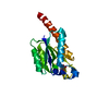
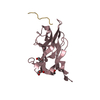
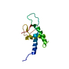
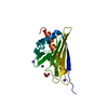
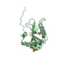
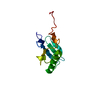
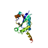
| Title | NMR structure of Rpa0253 from Rhodopseudomonas palustris. Northeast structural genomics consortium target RpR3 | ||||||
 Components Components | Hypothetical protein Rpa0253 | ||||||
 Keywords Keywords | STRUCTURAL GENOMICS / UNKNOWN FUNCTION / alpha-beta / RpR3 / NESG / Rpa0253 / COG2947 / PSI / Protein Structure Initiative / Northeast Structural Genomics Consortium | ||||||
| Function / homology |  Function and homology information Function and homology information: / : / EVE domain / EVE domain / ph1033 like fold / ph1033 like domains / PUA-like superfamily / Roll / Alpha Beta Similarity search - Domain/homology | ||||||
| Biological species |  Rhodopseudomonas palustris (phototrophic) Rhodopseudomonas palustris (phototrophic) | ||||||
| Method | SOLUTION NMR / distance geometry, simulated annealing, torsion angle dynamics, cns water refinement | ||||||
 Authors Authors | Ramelot, T.A. / Cort, J.R. / Conover, K. / Chen, Y. / Ma, L.C. / Ciano, M. / Xiao, R. / Acton, T.B. / Montelione, G.T. / Kennedy, M.A. / Northeast Structural Genomics Consortium (NESG) | ||||||
 Citation Citation | Journal: Proteins / Year: 2009 Title: Structural genomics reveals EVE as a new ASCH/PUA-related domain. Authors: Bertonati, C. / Punta, M. / Fischer, M. / Yachdav, G. / Forouhar, F. / Zhou, W. / Kuzin, A.P. / Seetharaman, J. / Abashidze, M. / Ramelot, T.A. / Kennedy, M.A. / Cort, J.R. / Belachew, A. / ...Authors: Bertonati, C. / Punta, M. / Fischer, M. / Yachdav, G. / Forouhar, F. / Zhou, W. / Kuzin, A.P. / Seetharaman, J. / Abashidze, M. / Ramelot, T.A. / Kennedy, M.A. / Cort, J.R. / Belachew, A. / Hunt, J.F. / Tong, L. / Montelione, G.T. / Rost, B. | ||||||
| History |
|
- Structure visualization
| Structure viewer | Molecule: MolmilJmol/JSmol |
|---|
- Downloads & links
-Download
| PDBx/mmCIF format | 2gbs.cif.gz | 883.3 KB | Display | PDBx/mmCIF format |
|---|---|---|---|---|
| PDB format | pdb2gbs.ent.gz | 743.5 KB | Display | PDB format |
| PDBx/mmJSON format | 2gbs.json.gz | Tree view | PDBx/mmJSON format | |
| Others |  Other downloads Other downloads |
-Validation report
| Arichive directory | https://data.pdbj.org/pub/pdb/validation_reports/gb/2gbsftp://data.pdbj.org/pub/pdb/validation_reports/gb/2gbs | HTTPS FTP |
|---|
-Related structure data
| Related structure data | 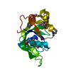 1zceC 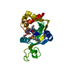 2eveC 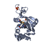 2g2xC C: citing same article ( |
|---|---|
| Similar structure data | |
| Other databases |


-Links
 PDBj
PDBj
- Assembly
| Deposited unit | 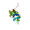
| |||||||||
|---|---|---|---|---|---|---|---|---|---|---|
| 1 |
| |||||||||
| NMR ensembles |
|
-Components
| #1: Protein | Mass: 16284.735 Da / Num. of mol.: 1 / Mutation: M1V Source method: isolated from a genetically manipulated source Source: (gene. exp.) Rhodopseudomonas palustris (phototrophic)Strain: CGA009 / Gene: rpa0253 / Production host: |
|---|
-Experimental details
-Experiment
| Experiment | Method: SOLUTION NMR | ||||||||||||||||||||
|---|---|---|---|---|---|---|---|---|---|---|---|---|---|---|---|---|---|---|---|---|---|
| NMR experiment |
|
- Sample preparation
| Details | Contents: 1 mM Rpa0253, U-15N, 13C; 20mM MES, 100mM NaCl, 5mM CaCl2, 10 mM DTT, 0.02% NaN3; 95% H2O, 5% D2O, pH 6.5 Solvent system: 95% H2O/5% D2O |
|---|---|
| Sample conditions | Ionic strength: 100mM NaCL / pH: 6.5 / Pressure: ambient / Temperature: 293 K |
-NMR measurement
| Radiation | Protocol: SINGLE WAVELENGTH / Monochromatic (M) / Laue (L): M / Scattering type: x-ray | |||||||||||||||
|---|---|---|---|---|---|---|---|---|---|---|---|---|---|---|---|---|
| Radiation wavelength | Relative weight: 1 | |||||||||||||||
| NMR spectrometer |
|
- Processing
| NMR software |
| ||||||||||||||||||||||||||||
|---|---|---|---|---|---|---|---|---|---|---|---|---|---|---|---|---|---|---|---|---|---|---|---|---|---|---|---|---|---|
| Refinement | Method: distance geometry, simulated annealing, torsion angle dynamics, cns water refinement Software ordinal: 1 Details: THE STRUCTURES ARE BASED ON A TOTAL OF 1690 RESTRAINTS. 1512 are NOE-DERIVED; SEQUENTIAL [(I-J)=1] = 201; MEDIUM RANGE [10.1 ANG = 0; AVERAGE RMS DISTANCE VIOLATION / CONSTRAINT = 0.001 ANG.; ...Details: THE STRUCTURES ARE BASED ON A TOTAL OF 1690 RESTRAINTS. 1512 are NOE-DERIVED; SEQUENTIAL [(I-J)=1] = 201; MEDIUM RANGE [1<(I-J)<5] = 411; LONG RANGE [(I-J)>=5] = 900; HYDROGEN BOND RESTRAINTS = 80 (2 PER H-BOND); NUMBER OF NOE RESTRAINTS PER RESIDUE = 11.0 (RESIDES 2-138); DIHEDRAL-ANGLE RESTRAINTS = 178 (89 PHI, 89 PSI); TOTAL NUMBER OF RESTRAINTS PER RESIDUE = 12.2 (RESIDES 1-138); NUMBER OF LONG RANGE RESTRAINTS PER RESIDUE = 6.5; NUMBER OF STRUCTURES COMPUTED = 20; NUMBER OF STRUCTURES USED = 20. AVERAGE DISTANCE VIOLATIONS >0.1 ANG = 0; AVERAGE RMS DISTANCE VIOLATION / CONSTRAINT = 0.001 ANG.; MAXIMUM DISTANCE VIOLATION 0.038. AVERAGE DIHEDRAL ANGLE VIOLATIONS: >1 DEG = 0; MAX DIHEDRAL ANGLE VIOLATION = 0.73; AVERAGE RMS ANGLE VIOLATION / CONSTRAINT = 0.03 DEG. RMSD VALUES: BACKBONE ATOMS (N,C,C', RESIDUES 2-136) = 0.57 ANG; ALL HEAVY ATOMS = 0.93 ANG; PROCHECK (RESIDUES 2-136): MOST FAVORED REGIONS = 90%; ADDITIONAL ALLOWED REGIONS = 9%; GENEROUSLY ALLOWED REGIONS = 0%; DISALLOWED REGIONS = 1%. THE 8 RESIDUE C-TERMINAL HIS TAG (LEHHHHHH) WAS INCLUDED IN THE STRUCTURE CALCULATION. | ||||||||||||||||||||||||||||
| NMR representative | Selection criteria: closest to the average | ||||||||||||||||||||||||||||
| NMR ensemble | Conformer selection criteria: all calculated structures submitted Conformers calculated total number: 20 / Conformers submitted total number: 20 |
 NMRPipe