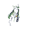
SHEET The sheet structure of this molecule is bifurcated. In order to represent this feature in the ...SHEET The sheet structure of this molecule is bifurcated. In order to represent this feature in the sheet report below, two sheets are defined. Strands 1,2 and 3 in sheet A and B are identical.
Mass: 11903.733 Da / Num. of mol.: 1 Source method: isolated from a genetically manipulated source Source: (gene. exp.) Escherichia coli (E. coli) / Strain: O157:H7 EDL933 / Gene: yggU / Plasmid: ER14-21 / Production host: Escherichia coli (E. coli) / Strain (production host): BL21pMgk / References: UniProt: Q8XCU6
-
Experimental details
-
Experiment
Experiment
Method: SOLUTION NMR
NMR experiment
Conditions-ID
Experiment-ID
Solution-ID
Type
1
1
1
3D 15N-NOESY
1
2
1
3D 13C-NOESY
1
3
1
3D aromatic 13C-NOESY
1
4
2
2D 15N,1H HSQC-J
1
5
1
2D15N,1HMEXICO
NMR details
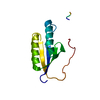
Text: The structure was determined using triple-resonance NMR spectroscopy. Automatic backbone resonance assignments were made using AUTOASSIGN. Automatic NOESY assignments as well as distance and ...Text: The structure was determined using triple-resonance NMR spectroscopy. Automatic backbone resonance assignments were made using AUTOASSIGN. Automatic NOESY assignments as well as distance and hydrogen bond restraints were determined using the AUTOSTRUCTURE program. Dihedral angle restraints were determined using HYPER and TALOS. Backbone conformations for residues 1-5, 25-26, 66, 101-108 are not well-defined [S(phi) + S(psi) < 1.8] in this solution NMR structure.
-
Sample preparation
Details
Solution-ID
Contents
Solvent system
1
1.0 mM ER14 U-15N,13C in 20mM MES, 50mM NaCl, 5mM DTT, pH 6.5
95% H2O/5% D2O
2
1.0 mM ER14 U-15N,13C in 20mM MES, 50mM NaCl, 5mM DTT, pH 6.5
95% H2O/5% D2O
Sample conditions
Ionic strength: 50 mM NaCl / pH: 6.5 / Pressure: ambient / Temperature: 293 K
Crystal grow
*PLUS
Method: other / Details: NMR
-
NMR measurement
Radiation
Protocol: SINGLE WAVELENGTH / Monochromatic (M) / Laue (L): M / Scattering type: x-ray
Radiation wavelength
Relative weight: 1
NMR spectrometer
Type
Manufacturer
Model
Field strength (MHz)
Spectrometer-ID
Varian INOVA
Varian
INOVA
500
1
Varian INOVA
Varian
INOVA
600
2
Varian INOVA
Varian
INOVA
750
3
-
Processing
NMR software
Name
Version
Developer
Classification
VNMR
6.1B
VARIANInc.
collection
NMRPipe
2.1
Delaglio, Bax
processing
Sparky
3.106
Goddard, Kneller
dataanalysis
AutoAssign
1.9
Moseley, Zimmerman, Montelione
dataanalysis
AutoStructure
1.1.2
Huang, Montelione
structuresolution
HYPER
2.7
Tejero, Montelione
structuresolution
TALOS
2.1
Cornilescu, Delaglio, Bax
structuresolution
DYANA
1.5
Guntert
refinement
X-PLOR
2.0.4 (NIH)
Brunger
refinement
PdbStat
3.27
Tejero, Montelione
dataanalysis
AutoStructure
1.1.2
Huang
refinement
Refinement
Method: torsion angle dynamics simulated annealing / Software ordinal: 1 Details: The structures are based on a total of 1419 conformationally-restricting NOE-derived distance restraints, 210 dihedral angle restraints, and 78 hydrogen bond restraints. Initial structure ...Details: The structures are based on a total of 1419 conformationally-restricting NOE-derived distance restraints, 210 dihedral angle restraints, and 78 hydrogen bond restraints. Initial structure determination was performed by torsion angle dynamics (DYANA). The final structures submitted were refined by simulated annealing (XPLOR).
NMR representative
Selection criteria: lowest energy
NMR ensemble
Conformer selection criteria: structures with the lowest energy Conformers calculated total number: 100 / Conformers submitted total number: 10
+
About Yorodumi
-
News
-
Feb 9, 2022. New format data for meta-information of EMDB entries
New format data for meta-information of EMDB entries
Version 3 of the EMDB header file is now the official format.
The previous official version 1.9 will be removed from the archive.
In the structure databanks used in Yorodumi, some data are registered as the other names, "COVID-19 virus" and "2019-nCoV". Here are the details of the virus and the list of structure data.
Jan 31, 2019. EMDB accession codes are about to change! (news from PDBe EMDB page)
EMDB accession codes are about to change! (news from PDBe EMDB page)
The allocation of 4 digits for EMDB accession codes will soon come to an end. Whilst these codes will remain in use, new EMDB accession codes will include an additional digit and will expand incrementally as the available range of codes is exhausted. The current 4-digit format prefixed with “EMD-” (i.e. EMD-XXXX) will advance to a 5-digit format (i.e. EMD-XXXXX), and so on. It is currently estimated that the 4-digit codes will be depleted around Spring 2019, at which point the 5-digit format will come into force.
The EM Navigator/Yorodumi systems omit the EMD- prefix.
Related info.:Q: What is EMD? / ID/Accession-code notation in Yorodumi/EM Navigator
Yorodumi is a browser for structure data from EMDB, PDB, SASBDB, etc.
This page is also the successor to EM Navigator detail page, and also detail information page/front-end page for Omokage search.
The word "yorodu" (or yorozu) is an old Japanese word meaning "ten thousand". "mi" (miru) is to see.
Related info.:EMDB / PDB / SASBDB / Comparison of 3 databanks / Yorodumi Search / Aug 31, 2016. New EM Navigator & Yorodumi / Yorodumi Papers / Jmol/JSmol / Function and homology information / Changes in new EM Navigator and Yorodumi
 Movie
Movie Controller

 Yorodumi
 Open data
 Basic information
 Components
 Keywords
 Function and homology information
 Authors
 Citation
 Structure visualization
 Downloads & links
 Other downloads
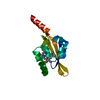


 PDBj
PDBj Assembly
 HSQC
HSQC Sample preparation
 Processing