[English] 日本語
Yorodumi
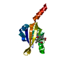
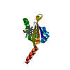
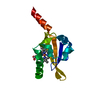
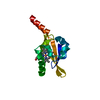
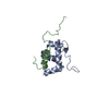
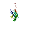
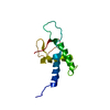
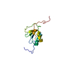
- PDB-1drm: CRYSTAL STRUCTURE OF THE LIGAND FREE BJFIXL HEME DOMAIN -

+
Open data


ID or keywords:

Loading...

-
Basic information

Entry
Database: PDB / ID: 1drm
TitleCRYSTAL STRUCTURE OF THE LIGAND FREE BJFIXL HEME DOMAIN
ComponentsSENSOR PROTEIN FIXL
KeywordsTRANSFERASE / FixL / heme domain / PAS family / two-component system / histidine kinase
Function / homology
Function and homology information


histidine phosphotransfer kinase activity / nitrogen fixation / histidine kinase / phosphorelay sensor kinase activity / regulation of DNA-templated transcription / ATP binding / metal ion binding
Similarity search - Function
PAS-associated, C-terminal / PAC domain profile. / His Kinase A (phospho-acceptor) domain / His Kinase A (phosphoacceptor) domain / PAC motif / Motif C-terminal to PAS motifs (likely to contribute to PAS structural domain) / Signal transduction histidine kinase, dimerisation/phosphoacceptor domain / Signal transduction histidine kinase, dimerisation/phosphoacceptor domain superfamily / PAS domain / Signal transduction histidine kinase-related protein, C-terminal ...PAS-associated, C-terminal / PAC domain profile. / His Kinase A (phospho-acceptor) domain / His Kinase A (phosphoacceptor) domain / PAC motif / Motif C-terminal to PAS motifs (likely to contribute to PAS structural domain) / Signal transduction histidine kinase, dimerisation/phosphoacceptor domain / Signal transduction histidine kinase, dimerisation/phosphoacceptor domain superfamily / PAS domain / Signal transduction histidine kinase-related protein, C-terminal / Histidine kinase domain / Histidine kinase domain profile. / Beta-Lactamase / PAS fold / PAS fold / PAS domain / PAS repeat profile. / PAS domain / Histidine kinase-, DNA gyrase B-, and HSP90-like ATPase / PAS domain superfamily / Histidine kinase-like ATPases / Histidine kinase/HSP90-like ATPase / Histidine kinase/HSP90-like ATPase superfamily / 2-Layer Sandwich / Alpha Beta
Similarity search - Domain/homology
PROTOPORPHYRIN IX CONTAINING FE / Sensor protein FixL
Similarity search - Component
Biological speciesBradyrhizobium japonicum (bacteria)
MethodX-RAY DIFFRACTION / SYNCHROTRON / Resolution: 2.4 Å
AuthorsGong, W. / Hao, B. / Mansy, S.S. / Gonzalez, G. / Gilles-Gonzalez, M.A. / Chan, M.K.
CitationJournal: Proc.Natl.Acad.Sci.USA / Year: 1998
Title: Structure of a biological oxygen sensor: a new mechanism for heme-driven signal transduction.
Authors: Gong, W. / Hao, B. / Mansy, S.S. / Gonzalez, G. / Gilles-Gonzalez, M.A. / Chan, M.K.
History
DepositionJan 6, 2000Deposition site: RCSB / Processing site: RCSB
SupersessionJan 24, 2000ID: 1BV6
Revision 1.0Jan 24, 2000Provider: repository / Type: Initial release
Revision 1.1Apr 27, 2008Group: Version format compliance
Revision 1.2Jul 13, 2011Group: Version format compliance
Revision 1.3Feb 7, 2024Group: Data collection / Database references / Derived calculations
Category: chem_comp_atom / chem_comp_bond ...chem_comp_atom / chem_comp_bond / database_2 / struct_conn / struct_ref_seq_dif / struct_site
Item: _database_2.pdbx_DOI / _database_2.pdbx_database_accession ..._database_2.pdbx_DOI / _database_2.pdbx_database_accession / _struct_conn.ptnr1_auth_comp_id / _struct_conn.ptnr1_auth_seq_id / _struct_conn.ptnr1_label_asym_id / _struct_conn.ptnr1_label_atom_id / _struct_conn.ptnr1_label_comp_id / _struct_conn.ptnr1_label_seq_id / _struct_conn.ptnr2_auth_comp_id / _struct_conn.ptnr2_auth_seq_id / _struct_conn.ptnr2_label_asym_id / _struct_conn.ptnr2_label_atom_id / _struct_conn.ptnr2_label_comp_id / _struct_conn.ptnr2_label_seq_id / _struct_ref_seq_dif.details / _struct_site.pdbx_auth_asym_id / _struct_site.pdbx_auth_comp_id / _struct_site.pdbx_auth_seq_id

-
Structure visualization

Structure viewerMolecule:
MolmilJmol/JSmol

Downloads & links

-
Assembly

Deposited unit
A: SENSOR PROTEIN FIXL
hetero molecules


Theoretical massNumber of molelcules
Total (without water)15,5362
Polymers14,9201
Non-polymers6161
Water1,47782
1


  • Idetical with deposited unit
  • defined by author
TypeNameSymmetry operationNumber
identity operation1_555x,y,z1
Unit cell
Length a, b, c (Å)128.800, 128.800, 58.900
Angle α, β, γ (deg.)90.00, 90.00, 120.00
Int Tables number155
Space group name H-MH32

-
Components

#1: Protein SENSOR PROTEIN FIXL /


Mass: 14919.858 Da / Num. of mol.: 1 / Fragment: HEME DOMAIN
Source method: isolated from a genetically manipulated source
Source: (gene. exp.) Bradyrhizobium japonicum (bacteria) / Production host: Escherichia coli (E. coli)
References: UniProt: P23222, Transferases; Transferring phosphorus-containing groups; Phosphotransferases with a nitrogenous group as acceptor
#2: Chemical ChemComp-HEM / PROTOPORPHYRIN IX CONTAINING FE / HEME / Heme B


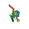
Mass: 616.487 Da / Num. of mol.: 1 / Source method: obtained synthetically / Formula: C34H32FeN4O4
#3: Water ChemComp-HOH / water / Water


Mass: 18.015 Da / Num. of mol.: 82 / Source method: isolated from a natural source / Formula: H2O

-
Experimental details

-
Experiment

ExperimentMethod: X-RAY DIFFRACTION / Number of used crystals: 1

-
Sample preparation

CrystalDensity Matthews: 3.15 Å3/Da / Density % sol: 60.95 %
Crystal growTemperature: 277 K / Method: vapor diffusion, hanging drop / pH: 7.5
Details: Sodium Chloride, MPD, HEPES, pH 7.5, VAPOR DIFFUSION, HANGING DROP, temperature 277K
Crystal grow
*PLUS
Temperature: 4 ℃ / Method: vapor diffusion
Components of the solutions
*PLUS
IDConc.Common nameCrystal-IDSol-IDChemical formula
14.5 M1reservoirNaCl
25 %MPD1reservoir
30.1 MHEPES1reservoir

-
Data collection

Diffraction
IDMean temperature (K)Crystal-ID
11001
21001
31001
41001
Diffraction source
SourceSiteBeamlineIDWavelength
SYNCHROTRONNSLS X4A11.7396
SYNCHROTRONNSLS X4A21.7379
SYNCHROTRONNSLS X4A31.6984
SYNCHROTRONNSLS X4A41.4938
DetectorDetector: CCD
RadiationProtocol: SINGLE WAVELENGTH / Monochromatic (M) / Laue (L): M / Scattering type: x-ray
Radiation wavelength
IDWavelength (Å)Relative weight
11.73961
21.73791
31.69841
41.49381
ReflectionResolution: 2.4→20 Å / Num. all: 31111 / Num. obs: 7104 / % possible obs: 97 % / Observed criterion σ(F): 0 / Observed criterion σ(I): 0 / Rmerge(I) obs: 0.076
Reflection shellResolution: 2.4→20 Å / Rmerge(I) obs: 0.221 / Num. unique all: 7104 / % possible all: 98
Reflection
*PLUS
Num. measured all: 31111
Reflection shell
*PLUS
% possible obs: 98 %

-
Processing

Software
NameVersionClassification
PHASESphasing
X-PLOR3.851refinement
DENZOdata reduction
SCALEPACKdata scaling
RefinementResolution: 2.4→20 Å / σ(F): 0 / σ(I): 0 / Stereochemistry target values: Engh & Huber / Details: Used weighted full matrix least squares procedure.
RfactorNum. reflection% reflectionSelection details
Rfree0.248 569 -RANDOM
Rwork0.199 ---
all0.199 31111 --
obs0.199 7104 97 %-
Refinement stepCycle: LAST / Resolution: 2.4→20 Å
ProteinNucleic acidLigandSolventTotal
Num. atoms938 0 43 82 1063
Refine LS restraints
Refine-IDTypeDev ideal
X-RAY DIFFRACTIONx_angle_deg1.675
X-RAY DIFFRACTIONx_bond_d0.014
Software
*PLUS
Name: X-PLOR / Version: 3.851 / Classification: refinement
Refinement
*PLUS
Solvent computation
*PLUS
Displacement parameters
*PLUS

+
About Yorodumi

-
News

-
Feb 9, 2022. New format data for meta-information of EMDB entries

New format data for meta-information of EMDB entries

  • Version 3 of the EMDB header file is now the official format.
  • The previous official version 1.9 will be removed from the archive.

Related info.:EMDB header

External links:wwPDB to switch to version 3 of the EMDB data model

-
Aug 12, 2020. Covid-19 info

Covid-19 info

URL: https://pdbj.org/emnavi/covid19.php

New page: Covid-19 featured information page in EM Navigator.

Related info.:Covid-19 info / Mar 5, 2020. Novel coronavirus structure data

+
Mar 5, 2020. Novel coronavirus structure data

Novel coronavirus structure data

Related info.:Yorodumi Speices / Aug 12, 2020. Covid-19 info

External links:COVID-19 featured content - PDBj / Molecule of the Month (242):Coronavirus Proteases

+
Jan 31, 2019. EMDB accession codes are about to change! (news from PDBe EMDB page)

EMDB accession codes are about to change! (news from PDBe EMDB page)

  • The allocation of 4 digits for EMDB accession codes will soon come to an end. Whilst these codes will remain in use, new EMDB accession codes will include an additional digit and will expand incrementally as the available range of codes is exhausted. The current 4-digit format prefixed with “EMD-” (i.e. EMD-XXXX) will advance to a 5-digit format (i.e. EMD-XXXXX), and so on. It is currently estimated that the 4-digit codes will be depleted around Spring 2019, at which point the 5-digit format will come into force.
  • The EM Navigator/Yorodumi systems omit the EMD- prefix.

Related info.:Q: What is EMD? / ID/Accession-code notation in Yorodumi/EM Navigator

External links:EMDB Accession Codes are Changing Soon! / Contact to PDBj

+
Jul 12, 2017. Major update of PDB

Major update of PDB

  • wwPDB released updated PDB data conforming to the new PDBx/mmCIF dictionary.
  • This is a major update changing the version number from 4 to 5, and with Remediation, in which all the entries are updated.
  • In this update, many items about electron microscopy experimental information are reorganized (e.g. em_software).
  • Now, EM Navigator and Yorodumi are based on the updated data.

External links:wwPDB Remediation / Enriched Model Files Conforming to OneDep Data Standards Now Available in the PDB FTP Archive

-
Yorodumi

Thousand views of thousand structures

  • Yorodumi is a browser for structure data from EMDB, PDB, SASBDB, etc.
  • This page is also the successor to EM Navigator detail page, and also detail information page/front-end page for Omokage search.
  • The word "yorodu" (or yorozu) is an old Japanese word meaning "ten thousand". "mi" (miru) is to see.

Related info.:EMDB / PDB / SASBDB / Comparison of 3 databanks / Yorodumi Search / Aug 31, 2016. New EM Navigator & Yorodumi / Yorodumi Papers / Jmol/JSmol / Function and homology information / Changes in new EM Navigator and Yorodumi

Read more