 Movie
Movie Controller

[English] 日本語
 Yorodumi
- PDB-2eve: X-Ray Crystal Structure of Protein PSPTO5229 from Pseudomonas syr... -
+ Open data

- Basic information
| Entry | Database: PDB / ID: 2eve | ||||||
|---|---|---|---|---|---|---|---|
| Title | X-Ray Crystal Structure of Protein PSPTO5229 from Pseudomonas syringae. Northeast Structural Genomics Consortium Target PsR62 | ||||||
 Components Components | hypothetical protein PSPTO5229 | ||||||
 Keywords Keywords | STRUCTURAL GENOMICS / UNKNOWN FUNCTION / alpha-beta protein / PSI / Protein Structure Initiative / Northeast Structural Genomics Consortium / NESG | ||||||
| Function / homology |  Function and homology information Function and homology information: / : / EVE domain / EVE domain / ph1033 like fold / ph1033 like domains / PUA-like superfamily / Roll / Alpha Beta Similarity search - Domain/homology | ||||||
| Biological species |  Pseudomonas syringae pv. tomato str. DC3000 (bacteria) Pseudomonas syringae pv. tomato str. DC3000 (bacteria) | ||||||
| Method |  X-RAY DIFFRACTION / SYNCHROTRON / SAD / Resolution: 1.6 Å X-RAY DIFFRACTION / SYNCHROTRON / SAD / Resolution: 1.6 Å | ||||||
 Authors Authors | Forouhar, F. / Zhou, W. / Belachew, A. / Jayaraman, S. / Ciao, M. / Xiao, R. / Acton, T.B. / Montelione, G.T. / Hunt, J.F. / Tong, L. / Northeast Structural Genomics Consortium (NESG) | ||||||
 Citation Citation | Journal: Proteins / Year: 2009 Title: Structural genomics reveals EVE as a new ASCH/PUA-related domain. Authors: Bertonati, C. / Punta, M. / Fischer, M. / Yachdav, G. / Forouhar, F. / Zhou, W. / Kuzin, A.P. / Seetharaman, J. / Abashidze, M. / Ramelot, T.A. / Kennedy, M.A. / Cort, J.R. / Belachew, A. / ...Authors: Bertonati, C. / Punta, M. / Fischer, M. / Yachdav, G. / Forouhar, F. / Zhou, W. / Kuzin, A.P. / Seetharaman, J. / Abashidze, M. / Ramelot, T.A. / Kennedy, M.A. / Cort, J.R. / Belachew, A. / Hunt, J.F. / Tong, L. / Montelione, G.T. / Rost, B. | ||||||
| History |
|
- Structure visualization
| Structure viewer | Molecule: MolmilJmol/JSmol |
|---|
- Downloads & links
-Download
| PDBx/mmCIF format | 2eve.cif.gz | 49.4 KB | Display | PDBx/mmCIF format |
|---|---|---|---|---|
| PDB format | pdb2eve.ent.gz | 33.8 KB | Display | PDB format |
| PDBx/mmJSON format | 2eve.json.gz | Tree view | PDBx/mmJSON format | |
| Others |  Other downloads Other downloads |
-Validation report
| Arichive directory | https://data.pdbj.org/pub/pdb/validation_reports/ev/2eveftp://data.pdbj.org/pub/pdb/validation_reports/ev/2eve | HTTPS FTP |
|---|
-Related structure data
| Related structure data | 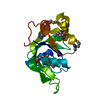 1zceC  2g2xC  2gbsC C: citing same article ( |
|---|---|
| Similar structure data | |
| Other databases |

-Links
 PDBj
PDBj
- Assembly
| Deposited unit | 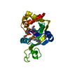
| ||||||||
|---|---|---|---|---|---|---|---|---|---|
| 1 |
| ||||||||
| Unit cell |
|
-Components
| #1: Protein | Mass: 18101.857 Da / Num. of mol.: 1 Source method: isolated from a genetically manipulated source Source: (gene. exp.) Pseudomonas syringae pv. tomato str. DC3000 (bacteria)Species: Pseudomonas syringae group genomosp. 3 / Strain: DC3000. / Gene: 1186914 / Plasmid: pET21+Magic / Production host: |
|---|---|
| #2: Chemical | ChemComp-MPO /   Mass: 209.263 Da / Num. of mol.: 1 / Source method: obtained synthetically / Formula: C7H15NO4S / Comment: pH buffer*YM Mass: 209.263 Da / Num. of mol.: 1 / Source method: obtained synthetically / Formula: C7H15NO4S / Comment: pH buffer*YM |
| #3: Chemical | ChemComp-EDO /   Mass: 62.068 Da / Num. of mol.: 1 / Source method: obtained synthetically / Formula: C2H6O2 Mass: 62.068 Da / Num. of mol.: 1 / Source method: obtained synthetically / Formula: C2H6O2 |
| #4: Chemical | ChemComp-144 /   Mass: 122.143 Da / Num. of mol.: 1 / Source method: obtained synthetically / Formula: C4H12NO3 Mass: 122.143 Da / Num. of mol.: 1 / Source method: obtained synthetically / Formula: C4H12NO3 |
| #5: Water | ChemComp-HOH /  Mass: 18.015 Da / Num. of mol.: 218 / Source method: isolated from a natural source / Formula: H2O Mass: 18.015 Da / Num. of mol.: 218 / Source method: isolated from a natural source / Formula: H2O |
| Has protein modification | Y |
-Experimental details
-Experiment
| Experiment | Method: X-RAY DIFFRACTION / Number of used crystals: 1 |
|---|
- Sample preparation
| Crystal | Density Matthews: 2.28 Å3/Da / Density % sol: 46.12 % / Description: The reported reflections include friedel pairs |
|---|---|
| Crystal grow | Temperature: 293 K / Method: vapor diffusion, hanging drop / pH: 7 Details: 0.5mM protein, 100mM MOPS, 30% PEG8000, 100mM NaH2PO4, 5mM DTT, pH 7, VAPOR DIFFUSION, HANGING DROP, temperature 293K |
-Data collection
| Diffraction | Mean temperature: 100 K |
|---|---|
| Diffraction source | Source: SYNCHROTRON / Site: NSLS  / Beamline: X4A / Wavelength: 0.9791 Å / Beamline: X4A / Wavelength: 0.9791 Å |
| Detector | Type: ADSC QUANTUM 4 / Detector: CCD / Date: Oct 27, 2005 / Details: mirrors. |
| Radiation | Monochromator: Si 111 CHANNEL / Protocol: SINGLE WAVELENGTH / Monochromatic (M) / Laue (L): M / Scattering type: x-ray |
| Radiation wavelength | Wavelength: 0.9791 Å / Relative weight: 1 |
| Reflection | Resolution: 1.55→30.71 Å / Num. all: 46260 / Num. obs: 46260 / % possible obs: 99.2 % / Observed criterion σ(F): 0 / Observed criterion σ(I): 0 / Redundancy: 14.1 % / Biso Wilson estimate: 15.2 Å2 / Rmerge(I) obs: 0.086 / Rsym value: 0.071 / Net I/σ(I): 23.75 |
| Reflection shell | Resolution: 1.55→1.61 Å / Redundancy: 14.2 % / Rmerge(I) obs: 0.394 / Mean I/σ(I) obs: 8.48 / Num. unique all: 2400 / Rsym value: 0.367 / % possible all: 98.6 |
- Processing
| Software |
| |||||||||||||||||||||||||
|---|---|---|---|---|---|---|---|---|---|---|---|---|---|---|---|---|---|---|---|---|---|---|---|---|---|---|
| Refinement | Method to determine structure: SAD / Resolution: 1.6→30.71 Å / Rfactor Rfree error: 0.004 / Data cutoff high absF: 586541.75 / Data cutoff low absF: 0 / Isotropic thermal model: OVERALL / Cross valid method: THROUGHOUT / σ(F): 2 / σ(I): 2 / Stereochemistry target values: Engh & Huber / Details: Friedel pairs have been used in refinement
| |||||||||||||||||||||||||
| Solvent computation | Solvent model: FLAT MODEL / Bsol: 42.8401 Å2 / ksol: 0.367001 e/Å3 | |||||||||||||||||||||||||
| Displacement parameters | Biso mean: 17.6 Å2
| |||||||||||||||||||||||||
| Refine analyze |
| |||||||||||||||||||||||||
| Refinement step | Cycle: LAST / Resolution: 1.6→30.71 Å
| |||||||||||||||||||||||||
| Refine LS restraints |
| |||||||||||||||||||||||||
| LS refinement shell | Resolution: 1.6→1.7 Å / Rfactor Rfree error: 0.009 / Total num. of bins used: 6
|
