 Movie
Movie Controller

[English] 日本語
 Yorodumi
- PDB-1czv: CRYSTAL STRUCTURE OF THE C2 DOMAIN OF HUMAN COAGULATION FACTOR V:... -
+ Open data

- Basic information
| Entry | Database: PDB / ID: 1czv | ||||||
|---|---|---|---|---|---|---|---|
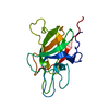
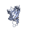
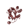
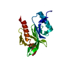
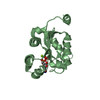
| Title | CRYSTAL STRUCTURE OF THE C2 DOMAIN OF HUMAN COAGULATION FACTOR V: DIMERIC CRYSTAL FORM | ||||||
 Components Components | PROTEIN (COAGULATION FACTOR V) | ||||||
 Keywords Keywords | BLOOD CLOTTING / COAGULATION / MEMBRANE-BINDING / DISCOIDIN FAMILY / CALCIUM-INDEPENDENT | ||||||
| Function / homology |  Function and homology information Function and homology informationresponse to vitamin K / platelet alpha granule / Cargo concentration in the ER / COPII-coated ER to Golgi transport vesicle / COPII-mediated vesicle transport / blood circulation / : / endoplasmic reticulum-Golgi intermediate compartment membrane / platelet alpha granule lumen / Post-translational protein phosphorylation ...response to vitamin K / platelet alpha granule / Cargo concentration in the ER / COPII-coated ER to Golgi transport vesicle / COPII-mediated vesicle transport / blood circulation / : / endoplasmic reticulum-Golgi intermediate compartment membrane / platelet alpha granule lumen / Post-translational protein phosphorylation / Regulation of Insulin-like Growth Factor (IGF) transport and uptake by Insulin-like Growth Factor Binding Proteins (IGFBPs) / blood coagulation / Platelet degranulation / extracellular vesicle / endoplasmic reticulum lumen / copper ion binding / : / extracellular region / membrane / plasma membrane Similarity search - Function | ||||||
| Biological species |  Homo sapiens (human) Homo sapiens (human) | ||||||
| Method |  X-RAY DIFFRACTION / MOLECULAR REPLACEMENT / Resolution: 2.4 Å X-RAY DIFFRACTION / MOLECULAR REPLACEMENT / Resolution: 2.4 Å | ||||||
 Authors Authors | Macedo-Ribeiro, S. / Bode, W. / Huber, R. / Kane, W.H. / Fuentes-Prior, P. | ||||||
 Citation Citation | Journal: Nature / Year: 1999 Title: Crystal structures of the membrane-binding C2 domain of human coagulation factor V. Authors: Macedo-Ribeiro, S. / Bode, W. / Huber, R. / Quinn-Allen, M.A. / Kim, S.W. / Ortel, T.L. / Bourenkov, G.P. / Bartunik, H.D. / Stubbs, M.T. / Kane, W.H. / Fuentes-Prior, P. | ||||||
| History |
|
- Structure visualization
| Structure viewer | Molecule: MolmilJmol/JSmol |
|---|
- Downloads & links
-Download
| PDBx/mmCIF format | 1czv.cif.gz | 76.6 KB | Display | PDBx/mmCIF format |
|---|---|---|---|---|
| PDB format | pdb1czv.ent.gz | 57.9 KB | Display | PDB format |
| PDBx/mmJSON format | 1czv.json.gz | Tree view | PDBx/mmJSON format | |
| Others |  Other downloads Other downloads |
-Validation report
| Arichive directory | https://data.pdbj.org/pub/pdb/validation_reports/cz/1czvftp://data.pdbj.org/pub/pdb/validation_reports/cz/1czv | HTTPS FTP |
|---|
-Related structure data
| Related structure data |  1czsSC  1cztC S: Starting model for refinement C: citing same article ( |
|---|---|
| Similar structure data |

-Links
 PDBj
PDBj


- Assembly
| Deposited unit | 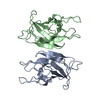
| ||||||||||
|---|---|---|---|---|---|---|---|---|---|---|---|
| 1 | 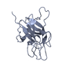
| ||||||||||
| 2 | 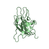
| ||||||||||
| Unit cell |
|
-Components
| #1: Protein | Mass: 18611.420 Da / Num. of mol.: 2 / Fragment: C2 DISCOIDIN-LIKE DOMAIN Source method: isolated from a genetically manipulated source Source: (gene. exp.) Homo sapiens (human) / Tissue: BLOOD / References: UniProt: P12259#2: Water | ChemComp-HOH / |  Mass: 18.015 Da / Num. of mol.: 88 / Source method: isolated from a natural source / Formula: H2O Mass: 18.015 Da / Num. of mol.: 88 / Source method: isolated from a natural source / Formula: H2OHas protein modification | Y | |
|---|
-Experimental details
-Experiment
| Experiment | Method: X-RAY DIFFRACTION / Number of used crystals: 1 |
|---|
- Sample preparation
| Crystal | Density Matthews: 2.48 Å3/Da / Density % sol: 45 % | ||||||||||||||||||||||||||||||
|---|---|---|---|---|---|---|---|---|---|---|---|---|---|---|---|---|---|---|---|---|---|---|---|---|---|---|---|---|---|---|---|
| Crystal grow | pH: 10 / Details: pH 10.00 | ||||||||||||||||||||||||||||||
| Crystal | *PLUS | ||||||||||||||||||||||||||||||
| Crystal grow | *PLUS Temperature: 20 ℃ / Method: vapor diffusion / PH range low: 10 / PH range high: 9.5 | ||||||||||||||||||||||||||||||
| Components of the solutions | *PLUS
|
-Data collection
| Diffraction | Mean temperature: 289 K |
|---|---|
| Diffraction source | Source: ROTATING ANODE / Type: RIGAKU / Wavelength: 1.5418 |
| Detector | Type: MARRESEARCH / Detector: IMAGE PLATE / Date: Jun 7, 1998 |
| Radiation | Protocol: SINGLE WAVELENGTH / Monochromatic (M) / Laue (L): M / Scattering type: x-ray |
| Radiation wavelength | Wavelength: 1.5418 Å / Relative weight: 1 |
| Reflection | Resolution: 2.4→24.6 Å / Num. obs: 14635 / % possible obs: 96.8 % / Observed criterion σ(I): 1 / Rmerge(I) obs: 0.094 |
| Reflection shell | Resolution: 2.4→2.51 Å / Rmerge(I) obs: 0.399 / % possible all: 90.7 |
| Reflection shell | *PLUS % possible obs: 90.7 % |
- Processing
| Software |
| ||||||||||||||||||||||||||||||||||||||||||||||||||||||||||||
|---|---|---|---|---|---|---|---|---|---|---|---|---|---|---|---|---|---|---|---|---|---|---|---|---|---|---|---|---|---|---|---|---|---|---|---|---|---|---|---|---|---|---|---|---|---|---|---|---|---|---|---|---|---|---|---|---|---|---|---|---|---|
| Refinement | Method to determine structure: MOLECULAR REPLACEMENT Starting model: 1CZS Resolution: 2.4→8 Å / σ(F): 1
| ||||||||||||||||||||||||||||||||||||||||||||||||||||||||||||
| Refinement step | Cycle: LAST / Resolution: 2.4→8 Å
| ||||||||||||||||||||||||||||||||||||||||||||||||||||||||||||
| Refine LS restraints |
| ||||||||||||||||||||||||||||||||||||||||||||||||||||||||||||
| Software | *PLUS Name: X-PLOR / Version: 3.851 / Classification: refinement | ||||||||||||||||||||||||||||||||||||||||||||||||||||||||||||
| Refinement | *PLUS Lowest resolution: 8 Å | ||||||||||||||||||||||||||||||||||||||||||||||||||||||||||||
| Solvent computation | *PLUS | ||||||||||||||||||||||||||||||||||||||||||||||||||||||||||||
| Displacement parameters | *PLUS |
