 Movie
Movie Controller

[English] 日本語
 Yorodumi
- PDB-2fzh: New Insights into Dihydrofolate Reductase Interactions: Analysis ... -
+ Open data

- Basic information
| Entry | Database: PDB / ID: 2fzh | ||||||
|---|---|---|---|---|---|---|---|
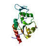
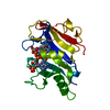
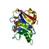
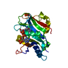
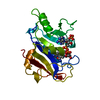
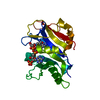
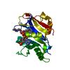
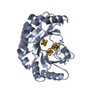
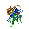
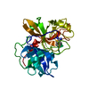
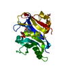
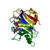
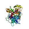
| Title | New Insights into Dihydrofolate Reductase Interactions: Analysis of Pneumocystis carinii and Mouse DHFR Complexes with NADPH and Two Highly Potent Trimethoprim Derivatives | ||||||
 Components Components | Dihydrofolate reductase | ||||||
 Keywords Keywords | OXIDOREDUCTASE / dihydrofolate reductase / Trimethoprim derivatives / ring stacking interactions | ||||||
| Function / homology |  Function and homology information Function and homology informationdihydrofolate metabolic process / dihydrofolate reductase / dihydrofolate reductase activity / folic acid metabolic process / tetrahydrofolate biosynthetic process / one-carbon metabolic process / NADP binding / mitochondrion Similarity search - Function | ||||||
| Biological species |  Pneumocystis carinii (fungus) Pneumocystis carinii (fungus) | ||||||
| Method |  X-RAY DIFFRACTION / MOLECULAR REPLACEMENT / Resolution: 2.1 Å X-RAY DIFFRACTION / MOLECULAR REPLACEMENT / Resolution: 2.1 Å | ||||||
 Authors Authors | Cody, V. / Pace, J. / Chisum, K. / Rosowsky, A. | ||||||
 Citation Citation | Journal: Proteins / Year: 2006 Title: New insights into DHFR interactions: analysis of Pneumocystis carinii and mouse DHFR complexes with NADPH and two highly potent 5-(omega-carboxy(alkyloxy) trimethoprim derivatives reveals ...Title: New insights into DHFR interactions: analysis of Pneumocystis carinii and mouse DHFR complexes with NADPH and two highly potent 5-(omega-carboxy(alkyloxy) trimethoprim derivatives reveals conformational correlations with activity and novel parallel ring stacking interactions. Authors: Cody, V. / Pace, J. / Chisum, K. / Rosowsky, A. | ||||||
| History |
|
- Structure visualization
| Structure viewer | Molecule: MolmilJmol/JSmol |
|---|
- Downloads & links
-Download
| PDBx/mmCIF format | 2fzh.cif.gz | 60.7 KB | Display | PDBx/mmCIF format |
|---|---|---|---|---|
| PDB format | pdb2fzh.ent.gz | 42.9 KB | Display | PDB format |
| PDBx/mmJSON format | 2fzh.json.gz | Tree view | PDBx/mmJSON format | |
| Others |  Other downloads Other downloads |
-Validation report
| Arichive directory | https://data.pdbj.org/pub/pdb/validation_reports/fz/2fzhftp://data.pdbj.org/pub/pdb/validation_reports/fz/2fzh | HTTPS FTP |
|---|
-Related structure data
| Related structure data | 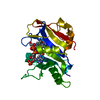 2fziC 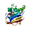 2fzjC  2cd2S S: Starting model for refinement C: citing same article ( |
|---|---|
| Similar structure data |


-Links
 PDBj
PDBj

- Assembly
| Deposited unit | 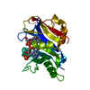
| ||||||||
|---|---|---|---|---|---|---|---|---|---|
| 1 |
| ||||||||
| Unit cell |
|
-Components
| #1: Protein | Mass: 23918.537 Da / Num. of mol.: 1 Source method: isolated from a genetically manipulated source Source: (gene. exp.) Pneumocystis carinii (fungus) / Plasmid: jm109 / Production host:  |
|---|---|
| #2: Chemical | ChemComp-NDP /   Mass: 745.421 Da / Num. of mol.: 1 / Source method: obtained synthetically / Formula: C21H30N7O17P3 Mass: 745.421 Da / Num. of mol.: 1 / Source method: obtained synthetically / Formula: C21H30N7O17P3 |
| #3: Chemical | ChemComp-DH1 /   Mass: 346.381 Da / Num. of mol.: 1 / Source method: obtained synthetically / Formula: C17H22N4O4 Mass: 346.381 Da / Num. of mol.: 1 / Source method: obtained synthetically / Formula: C17H22N4O4 |
| #4: Water | ChemComp-HOH /  Mass: 18.015 Da / Num. of mol.: 94 / Source method: isolated from a natural source / Formula: H2O Mass: 18.015 Da / Num. of mol.: 94 / Source method: isolated from a natural source / Formula: H2O |
-Experimental details
-Experiment
| Experiment | Method: X-RAY DIFFRACTION / Number of used crystals: 1 |
|---|
- Sample preparation
| Crystal | Density Matthews: 2.01 Å3/Da / Density % sol: 38.71 % |
|---|---|
| Crystal grow | Temperature: 298 K / Method: vapor diffusion, hanging drop / pH: 6 Details: 33% PEG 2K, 46 mM MES, 100 mM KCl, pH 6.0, VAPOR DIFFUSION, HANGING DROP, temperature 298K |
-Data collection
| Diffraction | Mean temperature: 200 K |
|---|---|
| Diffraction source | Source: ROTATING ANODE / Type: RIGAKU RU200 / Wavelength: 1.5418 Å |
| Detector | Type: RIGAKU RAXIS IV / Detector: IMAGE PLATE / Date: Sep 12, 2003 / Details: mirrors |
| Radiation | Monochromator: graphite / Protocol: SINGLE WAVELENGTH / Monochromatic (M) / Laue (L): M / Scattering type: x-ray |
| Radiation wavelength | Wavelength: 1.5418 Å / Relative weight: 1 |
| Reflection | Resolution: 2→25 Å / Num. all: 12754 / Num. obs: 11273 / % possible obs: 98.4 % / Observed criterion σ(F): 2 / Observed criterion σ(I): 2 / Redundancy: 3.2 % / Biso Wilson estimate: 14.6 Å2 / Rmerge(I) obs: 0.111 / Net I/σ(I): 16.5 |
| Reflection shell | Resolution: 2→2.07 Å / % possible all: 87.2 |
- Processing
| Software |
| |||||||||||||||||||||||||
|---|---|---|---|---|---|---|---|---|---|---|---|---|---|---|---|---|---|---|---|---|---|---|---|---|---|---|
| Refinement | Method to determine structure: MOLECULAR REPLACEMENT Starting model: PDB ENTRY 2cd2 Resolution: 2.1→25 Å / Cross valid method: THROUGHOUT / σ(F): 2 / Stereochemistry target values: Engh & Huber / Details: waters picked at level greater than 3.0
| |||||||||||||||||||||||||
| Displacement parameters | Biso mean: 42.4 Å2 | |||||||||||||||||||||||||
| Refinement step | Cycle: LAST / Resolution: 2.1→25 Å
| |||||||||||||||||||||||||
| Refine LS restraints |
| |||||||||||||||||||||||||
| Xplor file |
|
