 Movie
Movie Controller

[English] 日本語
 Yorodumi
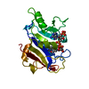
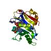
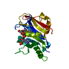
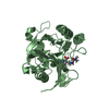
- PDB-1e26: Design, Synthesis and X-ray Crystal Structure of a Potent Dual In... -
+ Open data

- Basic information
| Entry | Database: PDB / ID: 1.0E+26 | ||||||
|---|---|---|---|---|---|---|---|
| Title | Design, Synthesis and X-ray Crystal Structure of a Potent Dual Inhibitor of Thymidylate Synthase and Dihydrofolate Reductase as an Antitumor Agent. | ||||||
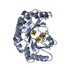
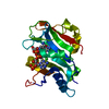
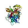
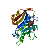
 Components Components | DIHYDROFOLATE REDUCTASE | ||||||
 Keywords Keywords | OXIDOREDUCTASE / DIHYDROFOLATE REDUCTASE / THYMIDYLATE SYNTHASE | ||||||
| Function / homology |  Function and homology information Function and homology informationdihydrofolate metabolic process / dihydrofolate reductase / dihydrofolate reductase activity / folic acid metabolic process / tetrahydrofolate biosynthetic process / one-carbon metabolic process / NADP binding / mitochondrion Similarity search - Function | ||||||
| Biological species |  PNEUMOCYSTIS CARINII (fungus) PNEUMOCYSTIS CARINII (fungus) | ||||||
| Method |  X-RAY DIFFRACTION / MOLECULAR REPLACEMENT / Resolution: 2 Å X-RAY DIFFRACTION / MOLECULAR REPLACEMENT / Resolution: 2 Å | ||||||
 Authors Authors | Gangjee, A. / Yu, J. / McGuire, J.J. / Cody, V. / Galitsky, N. / Kisliuk, R.L. / Queener, S.F. | ||||||
 Citation Citation | Journal: Biochemistry / Year: 1999 Title: Ligand-Induced Conformational Changes in the Crystal Structures of Pneumocystis Carinii Dihydrofolate Reductase Complexes with Folate and Nadp+ Authors: Cody, V. / Galitsky, N. / Rak, D. / Luft, J.R. / Pangborn, W. / Queener, S.F. | ||||||
| History |
| ||||||
| Remark 700 | SHEET THE SHEET STRUCTURE OF THIS MOLECULE IS BIFURCATED. IN ORDER TO REPRESENT THIS FEATURE IN ... SHEET THE SHEET STRUCTURE OF THIS MOLECULE IS BIFURCATED. IN ORDER TO REPRESENT THIS FEATURE IN THE SHEET RECORDS BELOW, TWO SHEETS ARE DEFINED. STRANDS 1 TO 7 SHEETS A AND A1 ARE IDENTICAL. |
- Structure visualization
| Structure viewer | Molecule: MolmilJmol/JSmol |
|---|
- Downloads & links
-Download
| PDBx/mmCIF format | 1e26.cif.gz | 61.3 KB | Display | PDBx/mmCIF format |
|---|---|---|---|---|
| PDB format | pdb1e26.ent.gz | 43.2 KB | Display | PDB format |
| PDBx/mmJSON format | 1e26.json.gz | Tree view | PDBx/mmJSON format | |
| Others |  Other downloads Other downloads |
-Validation report
| Arichive directory | https://data.pdbj.org/pub/pdb/validation_reports/e2/1e26ftp://data.pdbj.org/pub/pdb/validation_reports/e2/1e26 | HTTPS FTP |
|---|
-Related structure data
| Related structure data |  1cd2C  2cd2SC  3cd2C  4cd2C S: Starting model for refinement C: citing same article ( |
|---|---|
| Similar structure data |


-Links
 PDBj
PDBj
- Assembly
| Deposited unit | 
| ||||||||
|---|---|---|---|---|---|---|---|---|---|
| 1 |
| ||||||||
| Unit cell |
|
-Components
| #1: Protein | Mass: 23918.537 Da / Num. of mol.: 1 Source method: isolated from a genetically manipulated source Details: COMPLEXED WITH NADPH AND INHIBITOR / Source: (gene. exp.) PNEUMOCYSTIS CARINII (fungus) / Gene: C-DNA P.CARINII DHFR / Plasmid: PT7-7 / Gene (production host): C-DNA P.CARINII DHFR / Production host:  |
|---|---|
| #2: Chemical | ChemComp-NAP /   Mass: 743.405 Da / Num. of mol.: 1 / Source method: obtained synthetically / Formula: C21H28N7O17P3 Mass: 743.405 Da / Num. of mol.: 1 / Source method: obtained synthetically / Formula: C21H28N7O17P3 |
| #3: Chemical | ChemComp-GPB / 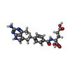  Mass: 425.438 Da / Num. of mol.: 1 / Source method: obtained synthetically / Formula: C21H23N5O5 Mass: 425.438 Da / Num. of mol.: 1 / Source method: obtained synthetically / Formula: C21H23N5O5 |
| #4: Water | ChemComp-HOH /  Mass: 18.015 Da / Num. of mol.: 101 / Source method: isolated from a natural source / Formula: H2O Mass: 18.015 Da / Num. of mol.: 101 / Source method: isolated from a natural source / Formula: H2O |
-Experimental details
-Experiment
| Experiment | Method: X-RAY DIFFRACTION / Number of used crystals: 1 |
|---|
- Sample preparation
| Crystal | Density Matthews: 1.97 Å3/Da / Density % sol: 48 % | ||||||||||||||||||||
|---|---|---|---|---|---|---|---|---|---|---|---|---|---|---|---|---|---|---|---|---|---|
| Crystal grow | Method: microbatch / pH: 6 Details: PROTEIN SOLUTION: WASHED AND CONCENTRATED 4 TUBES PCDHFR USING A CENTRICON 10 50 MM MES, 100MM KCL, B-NADPH, PH 6.0, A280/340 RATIO OF 24. WASHED WITH 50MM MES, 100MM KCL, PH 6.0, WASHED ...Details: PROTEIN SOLUTION: WASHED AND CONCENTRATED 4 TUBES PCDHFR USING A CENTRICON 10 50 MM MES, 100MM KCL, B-NADPH, PH 6.0, A280/340 RATIO OF 24. WASHED WITH 50MM MES, 100MM KCL, PH 6.0, WASHED WITH 50MM MES, 100MM KCL, PH 6.0 TO A CONCENTRATION OF 6.5 MG/ML. RESERVOIR SOLUTION: 50% PEG2000MME, 50MM MES, 100MM, PH 6.0 SET UP MICROBATCH POLYTHERMAL GRADIENT EXPERIMENTS IN MICROPETS. DROPS WERE COMPOSED OF: 2.5 MICROLITERS PROTEIN SOLUTION 2.0 MICROLITERS PROTEIN SOLUTION 0.5 MICROLITERS 50MM MES, 100MM KCL, PH 6.0. MICROPETS WERE LEFT TO EQUILIBRATE ON THE POLYTHERMAL GRADIENT. | ||||||||||||||||||||
| Crystal grow | *PLUS Temperature: 4 ℃ / pH: 6 / Method: unknown | ||||||||||||||||||||
| Components of the solutions | *PLUS
|
-Data collection
| Diffraction | Mean temperature: 293 K |
|---|---|
| Diffraction source | Source: ROTATING ANODE / Type: RIGAKU RU200HB / Wavelength: 1.5418 |
| Detector | Type: RIGAKU RAXIS IIC / Detector: IMAGE PLATE / Date: Jan 15, 1999 |
| Radiation | Protocol: SINGLE WAVELENGTH / Monochromatic (M) / Laue (L): M / Scattering type: x-ray |
| Radiation wavelength | Wavelength: 1.5418 Å / Relative weight: 1 |
| Reflection | Resolution: 2→100 Å / Num. obs: 11919 / % possible obs: 99.4 % / Observed criterion σ(I): 2 / Redundancy: 2.6 % / Rmerge(I) obs: 0.054 |
| Reflection | *PLUS Highest resolution: 2 Å / Lowest resolution: 50 Å / Redundancy: 2.6 % / Rmerge(I) obs: 0.049 |
| Reflection shell | *PLUS Highest resolution: 2 Å / Lowest resolution: 2.1 Å / % possible obs: 99.4 % |
- Processing
| Software |
| ||||||||||||||||||||||||||||||||||||||||||||||||||||||||||||
|---|---|---|---|---|---|---|---|---|---|---|---|---|---|---|---|---|---|---|---|---|---|---|---|---|---|---|---|---|---|---|---|---|---|---|---|---|---|---|---|---|---|---|---|---|---|---|---|---|---|---|---|---|---|---|---|---|---|---|---|---|---|
| Refinement | Method to determine structure: MOLECULAR REPLACEMENT Starting model: 2CD2 Resolution: 2→100 Å / Cross valid method: THROUGHOUT / σ(F): 0 Details: THE C-TERMINAL MET RESIDUE WAS NOT SEEN IN THE DENSITY MAPS
| ||||||||||||||||||||||||||||||||||||||||||||||||||||||||||||
| Refinement step | Cycle: LAST / Resolution: 2→100 Å
| ||||||||||||||||||||||||||||||||||||||||||||||||||||||||||||
| Refine LS restraints |
| ||||||||||||||||||||||||||||||||||||||||||||||||||||||||||||
| Software | *PLUS Name: CNS_SOLVE / Classification: refinement | ||||||||||||||||||||||||||||||||||||||||||||||||||||||||||||
| Refinement | *PLUS Lowest resolution: 50 Å / Num. reflection obs: 11919 / Rfactor obs: 0.223 / Rfactor Rfree: 0.297 / Rfactor Rwork: 0.223 | ||||||||||||||||||||||||||||||||||||||||||||||||||||||||||||
| Solvent computation | *PLUS | ||||||||||||||||||||||||||||||||||||||||||||||||||||||||||||
| Displacement parameters | *PLUS | ||||||||||||||||||||||||||||||||||||||||||||||||||||||||||||
| Refine LS restraints | *PLUS
|
