 Movie
Movie Controller

[English] 日本語
 Yorodumi
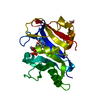
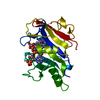
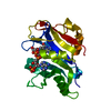
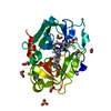
- PDB-1vj3: STRUCTURAL STUDIES ON BIO-ACTIVE COMPOUNDS. CRYSTAL STRUCTURE AND... -
+ Open data

- Basic information
| Entry | Database: PDB / ID: 1vj3 | |||||||||
|---|---|---|---|---|---|---|---|---|---|---|
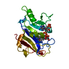
| Title | STRUCTURAL STUDIES ON BIO-ACTIVE COMPOUNDS. CRYSTAL STRUCTURE AND MOLECULAR MODELING STUDIES ON THE PNEUMOCYSTIS CARINII DIHYDROFOLATE REDUCTASE COFACTOR COMPLEX WITH TAB, A HIGHLY SELECTIVE ANTIFOLATE. | |||||||||
 Components Components | DIHYDROFOLATE REDUCTASE | |||||||||
 Keywords Keywords | OXIDOREDUCTASE / TAB / NADPH / MOLECULAR MODELING | |||||||||
| Function / homology |  Function and homology information Function and homology informationdihydrofolate metabolic process / dihydrofolate reductase / dihydrofolate reductase activity / folic acid metabolic process / tetrahydrofolate biosynthetic process / one-carbon metabolic process / NADP binding / mitochondrion Similarity search - Function | |||||||||
| Biological species |  Pneumocystis carinii (fungus) Pneumocystis carinii (fungus) | |||||||||
| Method |  X-RAY DIFFRACTION / Resolution: 2.1 Å X-RAY DIFFRACTION / Resolution: 2.1 Å | |||||||||
 Authors Authors | Cody, V. / Galitsky, N. / Rak, D. / Luft, J.R. / Queener, S.F. / Laughton, C.A. / Malcolm, F.G. | |||||||||
 Citation Citation | Journal: Biochemistry / Year: 2000 Title: Structural studies on bioactive compounds. 30. Crystal structure and molecular modeling studies on the Pneumocystis carinii dihydrofolate reductase cofactor complex with TAB, a highly selective antifolate. Authors: Cody, V. / Chan, D. / Galitsky, N. / Rak, D. / Luft, J.R. / Pangborn, W. / Queener, S.F. / Laughton, C.A. / Stevens, M.F. #1: Journal: Biochemistry / Year: 1999Title: Ligand-Induced Conformational Changes in the Crystal Structures of Pneumocystis Carinii Dihydrofolate Reductase Complexes with Folate and Nadp+ Authors: Cody, V. / Galitsky, N. / Rak, D. / Luft, J.R. / Pangborn, W. / Queener, S.F. #2: Journal: Structure / Year: 1994Title: The Structure of Pneumocystis Carinii Dihydrofolate Reductase to 1.9 A Resolution Authors: Champness, J.N. / Achari, A. / Ballantine, S.P. / Bryant, P.K. / Delves, C.J. / Stammers, D.K. | |||||||||
| History |
|
- Structure visualization
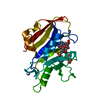
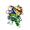
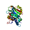
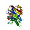
| Structure viewer | Molecule: MolmilJmol/JSmol |
|---|
- Downloads & links
-Download
| PDBx/mmCIF format | 1vj3.cif.gz | 61.1 KB | Display | PDBx/mmCIF format |
|---|---|---|---|---|
| PDB format | pdb1vj3.ent.gz | 43.1 KB | Display | PDB format |
| PDBx/mmJSON format | 1vj3.json.gz | Tree view | PDBx/mmJSON format | |
| Others |  Other downloads Other downloads |
-Validation report
| Arichive directory | https://data.pdbj.org/pub/pdb/validation_reports/vj/1vj3ftp://data.pdbj.org/pub/pdb/validation_reports/vj/1vj3 | HTTPS FTP |
|---|
-Related structure data
| Similar structure data |
|---|

-Links
 PDBj
PDBj

- Assembly
| Deposited unit | 
| ||||||||
|---|---|---|---|---|---|---|---|---|---|
| 1 |
| ||||||||
| Unit cell |
| ||||||||
| Atom site foot note | 1: CIS PROLINE - PRO 71 OMEGA = 0.02 2: CIS GLYCINE - GLY 125 OMEGA = 0.07 PEPTIDE BOND DEVIATES SIGNIFICANTLY FROM TRANS CONFORMATION |
-Components
| #1: Protein | Mass: 23787.340 Da / Num. of mol.: 1 Source method: isolated from a genetically manipulated source Source: (gene. exp.) Pneumocystis carinii (fungus) / Gene: C-DNA P.CARINII DHFR / Plasmid: PT7-7 / Production host:  |
|---|---|
| #2: Chemical | ChemComp-NDP /   Mass: 745.421 Da / Num. of mol.: 1 / Source method: obtained synthetically / Formula: C21H30N7O17P3 Mass: 745.421 Da / Num. of mol.: 1 / Source method: obtained synthetically / Formula: C21H30N7O17P3 |
| #3: Chemical | ChemComp-TAB /   Mass: 467.951 Da / Num. of mol.: 1 / Source method: obtained synthetically / Formula: C23H26ClN7O2 Mass: 467.951 Da / Num. of mol.: 1 / Source method: obtained synthetically / Formula: C23H26ClN7O2 |
| #4: Water | ChemComp-HOH /  Mass: 18.015 Da / Num. of mol.: 75 / Source method: isolated from a natural source / Formula: H2O Mass: 18.015 Da / Num. of mol.: 75 / Source method: isolated from a natural source / Formula: H2O |
-Experimental details
-Experiment
| Experiment | Method: X-RAY DIFFRACTION / Number of used crystals: 1 |
|---|
- Sample preparation
| Crystal | Density Matthews: 2.08 Å3/Da / Density % sol: 48 % | |||||||||||||||||||||||||||||||||||
|---|---|---|---|---|---|---|---|---|---|---|---|---|---|---|---|---|---|---|---|---|---|---|---|---|---|---|---|---|---|---|---|---|---|---|---|---|
| Crystal grow | pH: 6 / Details: pH 6.00 | |||||||||||||||||||||||||||||||||||
| Crystal grow | *PLUS Temperature: 4 ℃ / pH: 6 / Method: unknown | |||||||||||||||||||||||||||||||||||
| Components of the solutions | *PLUS
|
-Data collection
| Diffraction | Mean temperature: 293 K |
|---|---|
| Diffraction source | Source: ROTATING ANODE / Type: RIGAKU RU200 / Wavelength: 1.5418 |
| Detector | Type: RIGAKU RAXIS IIC / Detector: IMAGE PLATE / Date: Feb 1, 1998 |
| Radiation | Monochromator: GRAPH / Protocol: SINGLE WAVELENGTH / Monochromatic (M) / Laue (L): M / Scattering type: x-ray |
| Radiation wavelength | Wavelength: 1.5418 Å / Relative weight: 1 |
| Reflection | Resolution: 2.1→8 Å / Num. obs: 10325 / % possible obs: 97.8 % / Observed criterion σ(I): 1 / Redundancy: 2.2 % / Biso Wilson estimate: 16.7 Å2 / Rmerge(I) obs: 0.064 / Net I/σ(I): 4.1 |
| Reflection shell | Resolution: 2.1→2.18 Å / Redundancy: 3.1 % / Rmerge(I) obs: 0.187 / % possible all: 96.9 |
| Reflection | *PLUS Highest resolution: 2.1 Å / Lowest resolution: 8 Å / Rmerge(I) obs: 0.069 |
| Reflection shell | *PLUS % possible obs: 97.8 % |
- Processing
| Software |
| ||||||||||||||||||||||||||||||||||||||||||||||||||||||||||||||||||||||||||||||||||||
|---|---|---|---|---|---|---|---|---|---|---|---|---|---|---|---|---|---|---|---|---|---|---|---|---|---|---|---|---|---|---|---|---|---|---|---|---|---|---|---|---|---|---|---|---|---|---|---|---|---|---|---|---|---|---|---|---|---|---|---|---|---|---|---|---|---|---|---|---|---|---|---|---|---|---|---|---|---|---|---|---|---|---|---|---|---|
| Refinement | Resolution: 2.1→8 Å / σ(F): 2
| ||||||||||||||||||||||||||||||||||||||||||||||||||||||||||||||||||||||||||||||||||||
| Displacement parameters | Biso mean: 24.34 Å2 | ||||||||||||||||||||||||||||||||||||||||||||||||||||||||||||||||||||||||||||||||||||
| Refinement step | Cycle: LAST / Resolution: 2.1→8 Å
| ||||||||||||||||||||||||||||||||||||||||||||||||||||||||||||||||||||||||||||||||||||
| Refine LS restraints |
| ||||||||||||||||||||||||||||||||||||||||||||||||||||||||||||||||||||||||||||||||||||
| Refinement | *PLUS Highest resolution: 2.1 Å / Rfactor obs: 0.19 | ||||||||||||||||||||||||||||||||||||||||||||||||||||||||||||||||||||||||||||||||||||
| Solvent computation | *PLUS | ||||||||||||||||||||||||||||||||||||||||||||||||||||||||||||||||||||||||||||||||||||
| Displacement parameters | *PLUS |
