 Movie
Movie Controller

[English] 日本語
 Yorodumi
- PDB-1klk: CRYSTAL STRUCTURE OF PNEUMOCYSTIS CARINII DIHYDROFOLATE REDUCTASE... -
+ Open data

- Basic information
| Entry | Database: PDB / ID: 1klk | ||||||
|---|---|---|---|---|---|---|---|
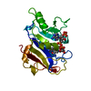
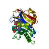
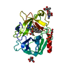
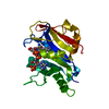
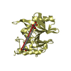
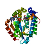
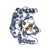
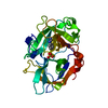
| Title | CRYSTAL STRUCTURE OF PNEUMOCYSTIS CARINII DIHYDROFOLATE REDUCTASE TERNARY COMPLEX WITH PT653 AND NADPH | ||||||
 Components Components | Dihydrofolate reductase | ||||||
 Keywords Keywords | OXIDOREDUCTASE / Structure-based design / antifolate / AIDS / protein-inhibitor complex / NADP / One-carbon metabolism | ||||||
| Function / homology |  Function and homology information Function and homology informationdihydrofolate metabolic process / dihydrofolate reductase / dihydrofolate reductase activity / folic acid metabolic process / tetrahydrofolate biosynthetic process / one-carbon metabolic process / NADP binding / mitochondrion Similarity search - Function | ||||||
| Biological species |  Pneumocystis carinii (fungus) Pneumocystis carinii (fungus) | ||||||
| Method |  X-RAY DIFFRACTION / MOLECULAR REPLACEMENT / Resolution: 2.3 Å X-RAY DIFFRACTION / MOLECULAR REPLACEMENT / Resolution: 2.3 Å | ||||||
 Authors Authors | Cody, V. / Galitsky, N. / Luft, J.R. / Pangborn, W. / Rosowsky, A. / Queener, S.F. | ||||||
 Citation Citation | Journal: Acta Crystallogr.,Sect.D / Year: 2002 Title: Structure-based enzyme inhibitor design: modeling studies and crystal structure analysis of Pneumocystis carinii dihydrofolate reductase ternary complex with PT653 and NADPH. Authors: Cody, V. / Galitsky, N. / Luft, J.R. / Pangborn, W. / Rosowsky, A. / Queener, S.F. #1: Journal: Biochemistry / Year: 2000Title: Structural Studies on Bioactive Compounds. 30. Crystal Structure and Molecular Modeling Studies on the Pneumocystis carinii Dihydrofolate Reductase Cofactor Complex with TAB, a Highly Selective Antifolate Authors: Cody, V. / Chan, D. / Galitsky, N. / Rak, D. / Luft, J.R. / Pangborn, W. / Queener, S.F. / Laughton, C.A. / Stevens, M.F.G. #2: Journal: Biochemistry / Year: 1999Title: Ligand-Induced Conformational Changes in the Crystal Structure of Pneumocystis carinii Dihydrofolate Reductase Complexes With Folate and NADP+ Authors: Cody, V. / Galitsky, N. / Rak, D. / Luft, J.R. / Pangborn, W. / Queener, S.F. | ||||||
| History |
|
- Structure visualization
| Structure viewer | Molecule: MolmilJmol/JSmol |
|---|
- Downloads & links
-Download
| PDBx/mmCIF format | 1klk.cif.gz | 57.5 KB | Display | PDBx/mmCIF format |
|---|---|---|---|---|
| PDB format | pdb1klk.ent.gz | 41.1 KB | Display | PDB format |
| PDBx/mmJSON format | 1klk.json.gz | Tree view | PDBx/mmJSON format | |
| Others |  Other downloads Other downloads |
-Validation report
| Arichive directory | https://data.pdbj.org/pub/pdb/validation_reports/kl/1klkftp://data.pdbj.org/pub/pdb/validation_reports/kl/1klk | HTTPS FTP |
|---|
-Related structure data
| Similar structure data |
|---|


-Links
 PDBj
PDBj

- Assembly
| Deposited unit | 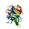
| ||||||||
|---|---|---|---|---|---|---|---|---|---|
| 1 |
| ||||||||
| Unit cell |
|
-Components
| #1: Protein | Mass: 23918.537 Da / Num. of mol.: 1 Source method: isolated from a genetically manipulated source Source: (gene. exp.) Pneumocystis carinii (fungus) / Genus: Rattus / Organ: lung / Plasmid: PT7-7 / Production host:  |
|---|---|
| #2: Chemical | ChemComp-PMD / [  Mass: 367.407 Da / Num. of mol.: 1 / Source method: obtained synthetically / Formula: C21H17N7 Mass: 367.407 Da / Num. of mol.: 1 / Source method: obtained synthetically / Formula: C21H17N7 |
| #3: Chemical | ChemComp-NDP /   Mass: 745.421 Da / Num. of mol.: 1 / Source method: obtained synthetically / Formula: C21H30N7O17P3 Mass: 745.421 Da / Num. of mol.: 1 / Source method: obtained synthetically / Formula: C21H30N7O17P3 |
| #4: Water | ChemComp-HOH /  Mass: 18.015 Da / Num. of mol.: 33 / Source method: isolated from a natural source / Formula: H2O Mass: 18.015 Da / Num. of mol.: 33 / Source method: isolated from a natural source / Formula: H2O |
-Experimental details
-Experiment
| Experiment | Method: X-RAY DIFFRACTION / Number of used crystals: 1 |
|---|
- Sample preparation
| Crystal | Density Matthews: 1.96 Å3/Da / Density % sol: 37.32 % | |||||||||||||||||||||||||||||||||||
|---|---|---|---|---|---|---|---|---|---|---|---|---|---|---|---|---|---|---|---|---|---|---|---|---|---|---|---|---|---|---|---|---|---|---|---|---|
| Crystal grow | Temperature: 298 K / Method: vapor diffusion / pH: 6 Details: PEG 2000MME, KCl, MES, pH 6.0, VAPOR DIFFUSION, temperature 298K | |||||||||||||||||||||||||||||||||||
| Crystal grow | *PLUS pH: 8.5 / Method: batch method | |||||||||||||||||||||||||||||||||||
| Components of the solutions | *PLUS
|
-Data collection
| Diffraction | Mean temperature: 298 K | ||||||||||||
|---|---|---|---|---|---|---|---|---|---|---|---|---|---|
| Diffraction source | Source: ROTATING ANODE / Type: RIGAKU RU200 / Wavelength: 1.5418,1.5621,1.7321 | ||||||||||||
| Detector | Type: RIGAKU RAXIS IV / Detector: IMAGE PLATE / Date: Oct 15, 1999 / Details: mirrors | ||||||||||||
| Radiation | Monochromator: graphite / Protocol: SINGLE WAVELENGTH / Monochromatic (M) / Laue (L): M / Scattering type: x-ray | ||||||||||||
| Radiation wavelength |
| ||||||||||||
| Reflection | Resolution: 2.3→25 Å / Num. all: 9597 / Num. obs: 5861 / % possible obs: 61.1 % / Observed criterion σ(F): 2 / Observed criterion σ(I): 1 | ||||||||||||
| Reflection shell | Resolution: 2.5→2.59 Å / Redundancy: 4.1 % / Rmerge(I) obs: 0.068 / Mean I/σ(I) obs: 23.4 / Num. unique all: 8889 / Rsym value: 0.234 / % possible all: 89.2 | ||||||||||||
| Reflection | *PLUS Rmerge(I) obs: 0.073 |
- Processing
| Software |
| ||||||||||||||||||||
|---|---|---|---|---|---|---|---|---|---|---|---|---|---|---|---|---|---|---|---|---|---|
| Refinement | Method to determine structure: MOLECULAR REPLACEMENT / Resolution: 2.3→8 Å / Isotropic thermal model: isotropic / Cross valid method: THROUGHOUT / σ(F): 0 / Stereochemistry target values: Engh & Huber
| ||||||||||||||||||||
| Displacement parameters | Biso mean: 44.4 Å2 | ||||||||||||||||||||
| Refinement step | Cycle: LAST / Resolution: 2.3→8 Å
| ||||||||||||||||||||
| Refine LS restraints |
| ||||||||||||||||||||
| LS refinement shell |
| ||||||||||||||||||||
| Refinement | *PLUS Highest resolution: 2.4 Å / Num. reflection obs: 5802 / Num. reflection Rfree: 634 | ||||||||||||||||||||
| Solvent computation | *PLUS | ||||||||||||||||||||
| Displacement parameters | *PLUS |
