 Movie
Movie Controller

[English] 日本語
 Yorodumi
- PDB-2fkc: Crystal Form I of Pre-Reactive Complex of Restriction Endonucleas... -
+ Open data

- Basic information
| Entry | Database: PDB / ID: 2fkc | ||||||
|---|---|---|---|---|---|---|---|
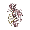
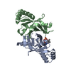
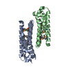
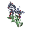
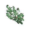
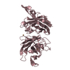
| Title | Crystal Form I of Pre-Reactive Complex of Restriction Endonuclease HinP1I with Cognate DNA and Calcium Ion | ||||||
 Components Components |
| ||||||
 Keywords Keywords | HYDROLASE/DNA / RESTRICTION ENDONUCLEASE / PROTEIN DIMERIZATON / DNA SUPERHELIX / PROTEIN-DNA-METAL ION COMPLEX / HYDROLASE-DNA COMPLEX | ||||||
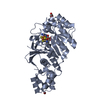
| Function / homology |  Function and homology information Function and homology information | ||||||
| Biological species |  Haemophilus influenzae (bacteria) Haemophilus influenzae (bacteria) | ||||||
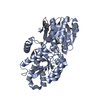
| Method |  X-RAY DIFFRACTION / SYNCHROTRON / MOLECULAR REPLACEMENT / Resolution: 2.39 Å X-RAY DIFFRACTION / SYNCHROTRON / MOLECULAR REPLACEMENT / Resolution: 2.39 Å | ||||||
 Authors Authors | Horton, J.R. | ||||||
 Citation Citation | Journal: Nucleic Acids Res. / Year: 2006 Title: DNA nicking by HinP1I endonuclease: bending, base flipping and minor groove expansion. Authors: Horton, J.R. / Zhang, X. / Maunus, R. / Yang, Z. / Wilson, G.G. / Roberts, R.J. / Cheng, X. #1: Journal: Nucleic Acids Res. / Year: 2005Title: Structure of HinP1I endonuclease reveals a striking similarity to the monomeric restriction enzyme MspI Authors: Yang, Z. / Horton, J.R. / Maunus, R. / Wilson, G.G. / Roberts, R.J. / Cheng, X. | ||||||
| History |
|
- Structure visualization
| Structure viewer | Molecule: MolmilJmol/JSmol |
|---|
- Downloads & links
-Download
| PDBx/mmCIF format | 2fkc.cif.gz | 135.8 KB | Display | PDBx/mmCIF format |
|---|---|---|---|---|
| PDB format | pdb2fkc.ent.gz | 102.8 KB | Display | PDB format |
| PDBx/mmJSON format | 2fkc.json.gz | Tree view | PDBx/mmJSON format | |
| Others |  Other downloads Other downloads |
-Validation report
| Arichive directory | https://data.pdbj.org/pub/pdb/validation_reports/fk/2fkcftp://data.pdbj.org/pub/pdb/validation_reports/fk/2fkc | HTTPS FTP |
|---|
-Related structure data
| Related structure data |  2fkhC  2fl3C  2flcC  1ynmS S: Starting model for refinement C: citing same article ( |
|---|---|
| Similar structure data |
-Links
 PDBj
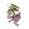
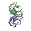
PDBj- Assembly
| Deposited unit | 
| ||||||||
|---|---|---|---|---|---|---|---|---|---|
| 1 | 
| ||||||||
| 2 | 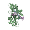
| ||||||||
| Unit cell |
|
-Components
| #1: DNA chain | Mass: 3045.992 Da / Num. of mol.: 4 / Source method: obtained synthetically / Details: Synthesized Self-Annealing Oligonucleotide #2: Protein | Mass: 28791.668 Da / Num. of mol.: 2 Source method: isolated from a genetically manipulated source Source: (gene. exp.) Haemophilus influenzae (bacteria) / Gene: hinP1IR / Plasmid: pUC19 / Production host: References: GenBank: 57116674, UniProt: Q5I6E6*PLUS, type II site-specific deoxyribonuclease #3: Chemical |   Mass: 40.078 Da / Num. of mol.: 3 / Source method: obtained synthetically / Formula: Ca Mass: 40.078 Da / Num. of mol.: 3 / Source method: obtained synthetically / Formula: Ca#4: Water | ChemComp-HOH / |  Mass: 18.015 Da / Num. of mol.: 44 / Source method: isolated from a natural source / Formula: H2O Mass: 18.015 Da / Num. of mol.: 44 / Source method: isolated from a natural source / Formula: H2O |
|---|
-Experimental details
-Experiment
| Experiment | Method: X-RAY DIFFRACTION / Number of used crystals: 1 |
|---|
- Sample preparation
| Crystal | Density Matthews: 2.95 Å3/Da / Density % sol: 58.37 % | ||||||||||||||||||||||||||||||||||||
|---|---|---|---|---|---|---|---|---|---|---|---|---|---|---|---|---|---|---|---|---|---|---|---|---|---|---|---|---|---|---|---|---|---|---|---|---|---|
| Crystal grow | Temperature: 289 K / Method: vapor diffusion, hanging drop / pH: 7.8 Details: 7.5%(v/v) ethanol, 1.5M NaCl, 100mM Bis-Tris propane pH 7.8, and 20mM CaCl2, VAPOR DIFFUSION, HANGING DROP, temperature 289K | ||||||||||||||||||||||||||||||||||||
| Components of the solutions |
|
-Data collection
| Diffraction | Mean temperature: 100 K |
|---|---|
| Diffraction source | Source: SYNCHROTRON / Site: APS  / Beamline: 22-ID / Wavelength: 0.97179 Å / Beamline: 22-ID / Wavelength: 0.97179 Å |
| Detector | Type: MARRESEARCH / Detector: CCD / Date: Feb 27, 2005 |
| Radiation | Monochromator: Si 220 / Protocol: SINGLE WAVELENGTH / Monochromatic (M) / Laue (L): M / Scattering type: x-ray |
| Radiation wavelength | Wavelength: 0.97179 Å / Relative weight: 1 |
| Reflection | Resolution: 2.39→19.89 Å / Num. all: 31659 / Num. obs: 31659 / % possible obs: 94.7 % / Observed criterion σ(F): -3 / Observed criterion σ(I): -3 / Redundancy: 7.1 % / Biso Wilson estimate: 47 Å2 / Rmerge(I) obs: 0.108 / Net I/σ(I): 19 |
| Reflection shell | Resolution: 2.39→2.48 Å / Redundancy: 7.1 % / Rmerge(I) obs: 0.368 / Num. unique all: 3183 / % possible all: 97.2 |
- Processing
| Software |
| |||||||||||||||||||||||||
|---|---|---|---|---|---|---|---|---|---|---|---|---|---|---|---|---|---|---|---|---|---|---|---|---|---|---|
| Refinement | Method to determine structure: MOLECULAR REPLACEMENT Starting model: pdb entry 1YNM Resolution: 2.39→19.89 Å / Isotropic thermal model: isotropic / Cross valid method: THROUGHOUT / σ(F): 0 / Stereochemistry target values: Engh & Huber
| |||||||||||||||||||||||||
| Displacement parameters | Biso mean: 59 Å2
| |||||||||||||||||||||||||
| Refine analyze |
| |||||||||||||||||||||||||
| Refinement step | Cycle: LAST / Resolution: 2.39→19.89 Å
| |||||||||||||||||||||||||
| Refine LS restraints |
|
