 Movie
Movie Controller

[English] 日本語
 Yorodumi
- PDB-5ahw: Crystal structure of universal stress protein MSMEG_3811 in compl... -
+ Open data

- Basic information
| Entry | Database: PDB / ID: 5ahw | ||||||
|---|---|---|---|---|---|---|---|
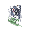
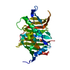
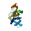
| Title | Crystal structure of universal stress protein MSMEG_3811 in complex with cAMP | ||||||
 Components Components | UNIVERSAL STRESS PROTEIN | ||||||
 Keywords Keywords | SIGNALING PROTEIN / RV1636 HOMOLOG / USP TYPE 1 HOMODIMER / WALKER A-LIKE MOTIF / ATP-BINDING MOTIF | ||||||
| Function / homology |  Function and homology information Function and homology information | ||||||
| Biological species |  MYCOBACTERIUM SMEGMATIS STR. MC2 155 (bacteria) MYCOBACTERIUM SMEGMATIS STR. MC2 155 (bacteria) | ||||||
| Method |  X-RAY DIFFRACTION / SYNCHROTRON / MOLECULAR REPLACEMENT / Resolution: 2.15 Å X-RAY DIFFRACTION / SYNCHROTRON / MOLECULAR REPLACEMENT / Resolution: 2.15 Å | ||||||
 Authors Authors | Adolph, R.S. / Kleinboelting, S. / Weyand, M. / Steegborn, C. | ||||||
 Citation Citation | Journal: J.Biol.Chem. / Year: 2015 Title: A Universal Stress Protein (Usp) in Mycobacteria Binds Camp Authors: Banerjee, A. / Adolph, R.S. / Gopalakrishnapai, J. / Kleinboelting, S. / Emmerich, C. / Steegborn, C. / Visweswariah, S.S. | ||||||
| History |
|
- Structure visualization
| Structure viewer | Molecule: MolmilJmol/JSmol |
|---|
- Downloads & links
-Download
| PDBx/mmCIF format | 5ahw.cif.gz | 301.3 KB | Display | PDBx/mmCIF format |
|---|---|---|---|---|
| PDB format | pdb5ahw.ent.gz | 248.6 KB | Display | PDB format |
| PDBx/mmJSON format | 5ahw.json.gz | Tree view | PDBx/mmJSON format | |
| Others |  Other downloads Other downloads |
-Validation report
| Arichive directory | https://data.pdbj.org/pub/pdb/validation_reports/ah/5ahwftp://data.pdbj.org/pub/pdb/validation_reports/ah/5ahw | HTTPS FTP |
|---|
-Related structure data
| Related structure data |  2z08S S: Starting model for refinement |
|---|---|
| Similar structure data |


-Links
 PDBj
PDBj


- Assembly
| Deposited unit | 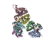
| |||||||||
|---|---|---|---|---|---|---|---|---|---|---|
| 1 | 
| |||||||||
| 2 | 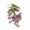
| |||||||||
| 3 | 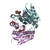
| |||||||||
| Unit cell |
| |||||||||
| Components on special symmetry positions |
|
-Components
-Protein , 1 types, 6 molecules ABCDEF
| #1: Protein | Mass: 15223.129 Da / Num. of mol.: 6 Source method: isolated from a genetically manipulated source Source: (gene. exp.) MYCOBACTERIUM SMEGMATIS STR. MC2 155 (bacteria)Plasmid: PPROEX-HTA / Production host: |
|---|
-Non-polymers , 5 types, 267 molecules 


| #2: Chemical | ChemComp-CMP /  Mass: 329.206 Da / Num. of mol.: 6 / Source method: obtained synthetically / Formula: C10H12N5O6P Mass: 329.206 Da / Num. of mol.: 6 / Source method: obtained synthetically / Formula: C10H12N5O6P#3: Chemical | ChemComp-SO4 /  Mass: 96.063 Da / Num. of mol.: 7 / Source method: obtained synthetically / Formula: SO4 Mass: 96.063 Da / Num. of mol.: 7 / Source method: obtained synthetically / Formula: SO4#4: Chemical |  Mass: 35.453 Da / Num. of mol.: 2 / Source method: obtained synthetically / Formula: Cl Mass: 35.453 Da / Num. of mol.: 2 / Source method: obtained synthetically / Formula: Cl#5: Chemical |  Mass: 424.569 Da / Num. of mol.: 3 / Source method: obtained synthetically / Formula: C21H44O8 Mass: 424.569 Da / Num. of mol.: 3 / Source method: obtained synthetically / Formula: C21H44O8#6: Water | ChemComp-HOH / | Mass: 18.015 Da / Num. of mol.: 249 / Source method: isolated from a natural source / Formula: H2O |
|---|
-Experimental details
-Experiment
| Experiment | Method: X-RAY DIFFRACTION / Number of used crystals: 1 |
|---|
- Sample preparation
| Crystal | Density Matthews: 2.04 Å3/Da / Density % sol: 39.9 % / Description: NONE |
|---|---|
| Crystal grow | pH: 6 Details: PROTEIN WAS COCRYSTALLIZED WITH 5 MM CAMP IN 0.1 M MES, PH 6.0; 1.9 M AMMONIUM SULFATE; 6 % (V/V) POLYPROPYLENGLYCOL 400. |
-Data collection
| Diffraction | Mean temperature: 100 K |
|---|---|
| Diffraction source | Source: SYNCHROTRON / Site: BESSY  / Beamline: 14.1 / Wavelength: 0.91841 / Beamline: 14.1 / Wavelength: 0.91841 |
| Detector | Type: DECTRIS PILATUS 6M / Detector: PIXEL / Date: Oct 23, 2013 / Details: COLLIMATOR |
| Radiation | Monochromator: SI111-DCM WITH SAGITTAL BENDER / Protocol: SINGLE WAVELENGTH / Monochromatic (M) / Laue (L): M / Scattering type: x-ray |
| Radiation wavelength | Wavelength: 0.91841 Å / Relative weight: 1 |
| Reflection | Resolution: 2.15→48.64 Å / Num. obs: 43084 / % possible obs: 98.8 % / Observed criterion σ(I): -3 / Redundancy: 4.5 % / Rmerge(I) obs: 0.08 / Net I/σ(I): 15.1 |
| Reflection shell | Resolution: 2.15→2.21 Å / Redundancy: 4.4 % / Rmerge(I) obs: 0.9 / Mean I/σ(I) obs: 1.8 / % possible all: 98.5 |
- Processing
| Software |
| ||||||||||||||||||||||||||||||||||||||||||||||||||||||||||||||||||||||||||||||||||||||||||||||||||||||||||||||||||||||||||||||||||||||||||||||||||||||||||||||||||||||||||||||||||||||
|---|---|---|---|---|---|---|---|---|---|---|---|---|---|---|---|---|---|---|---|---|---|---|---|---|---|---|---|---|---|---|---|---|---|---|---|---|---|---|---|---|---|---|---|---|---|---|---|---|---|---|---|---|---|---|---|---|---|---|---|---|---|---|---|---|---|---|---|---|---|---|---|---|---|---|---|---|---|---|---|---|---|---|---|---|---|---|---|---|---|---|---|---|---|---|---|---|---|---|---|---|---|---|---|---|---|---|---|---|---|---|---|---|---|---|---|---|---|---|---|---|---|---|---|---|---|---|---|---|---|---|---|---|---|---|---|---|---|---|---|---|---|---|---|---|---|---|---|---|---|---|---|---|---|---|---|---|---|---|---|---|---|---|---|---|---|---|---|---|---|---|---|---|---|---|---|---|---|---|---|---|---|---|---|
| Refinement | Method to determine structure: MOLECULAR REPLACEMENT Starting model: PDB ENTRY 2Z08 Resolution: 2.15→48.64 Å / Cor.coef. Fo:Fc: 0.966 / Cor.coef. Fo:Fc free: 0.94 / SU B: 12.713 / SU ML: 0.168 / Cross valid method: THROUGHOUT / ESU R: 0.237 / ESU R Free: 0.199 / Stereochemistry target values: MAXIMUM LIKELIHOOD / Details: HYDROGENS HAVE BEEN ADDED IN THE RIDING POSITIONS
| ||||||||||||||||||||||||||||||||||||||||||||||||||||||||||||||||||||||||||||||||||||||||||||||||||||||||||||||||||||||||||||||||||||||||||||||||||||||||||||||||||||||||||||||||||||||
| Solvent computation | Ion probe radii: 0.8 Å / Shrinkage radii: 0.8 Å / VDW probe radii: 1.2 Å / Solvent model: MASK | ||||||||||||||||||||||||||||||||||||||||||||||||||||||||||||||||||||||||||||||||||||||||||||||||||||||||||||||||||||||||||||||||||||||||||||||||||||||||||||||||||||||||||||||||||||||
| Refinement step | Cycle: LAST / Resolution: 2.15→48.64 Å
| ||||||||||||||||||||||||||||||||||||||||||||||||||||||||||||||||||||||||||||||||||||||||||||||||||||||||||||||||||||||||||||||||||||||||||||||||||||||||||||||||||||||||||||||||||||||
| Refine LS restraints |
|