 Movie
Movie Controller

[English] 日本語
 Yorodumi
- PDB-4qvb: Mycobacterium tuberculosis protein Rv1155 in complex with co-enzy... -
+ Open data

- Basic information
| Entry | Database: PDB / ID: 4qvb | ||||||
|---|---|---|---|---|---|---|---|
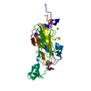
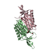
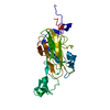
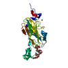
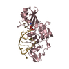
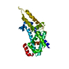
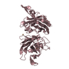
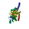
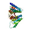
| Title | Mycobacterium tuberculosis protein Rv1155 in complex with co-enzyme F420 | ||||||
 Components Components | Rv1155 protein | ||||||
 Keywords Keywords | OXIDOREDUCTASE | ||||||
| Function / homology |  Function and homology information Function and homology informationvitamin B6 metabolic process / coenzyme F420 binding / oxidoreductase activity, acting on the CH-CH group of donors / Oxidoreductases / FMN binding / pyridoxal phosphate binding / protein homodimerization activity Similarity search - Function | ||||||
| Biological species |   Mycobacterium tuberculosis (bacteria) Mycobacterium tuberculosis (bacteria) | ||||||
| Method |  X-RAY DIFFRACTION / SYNCHROTRON / MOLECULAR REPLACEMENT / Resolution: 2.3 Å X-RAY DIFFRACTION / SYNCHROTRON / MOLECULAR REPLACEMENT / Resolution: 2.3 Å | ||||||
 Authors Authors | Mashalidis, E.H. / Gittis, A.G. / Tomczak, A. / Abell, C. / Barry III, C.E. / Garboczi, D.N. | ||||||
 Citation Citation | Journal: Protein Sci. / Year: 2015 Title: Molecular insights into the binding of coenzyme F420 to the conserved protein Rv1155 from Mycobacterium tuberculosis. Authors: Mashalidis, E.H. / Gittis, A.G. / Tomczak, A. / Abell, C. / Barry, C.E. / Garboczi, D.N. | ||||||
| History |
|
- Structure visualization
| Structure viewer | Molecule: MolmilJmol/JSmol |
|---|
- Downloads & links
-Download
| PDBx/mmCIF format | 4qvb.cif.gz | 73.3 KB | Display | PDBx/mmCIF format |
|---|---|---|---|---|
| PDB format | pdb4qvb.ent.gz | 53.9 KB | Display | PDB format |
| PDBx/mmJSON format | 4qvb.json.gz | Tree view | PDBx/mmJSON format | |
| Others |  Other downloads Other downloads |
-Validation report
| Arichive directory | https://data.pdbj.org/pub/pdb/validation_reports/qv/4qvbftp://data.pdbj.org/pub/pdb/validation_reports/qv/4qvb | HTTPS FTP |
|---|
-Related structure data
| Similar structure data |
|---|

-Links
 PDBj
PDBj- Assembly
| Deposited unit | 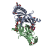
| ||||||||
|---|---|---|---|---|---|---|---|---|---|
| 1 |
| ||||||||
| Unit cell |
|
-Components
-Protein , 1 types, 2 molecules AB
| #1: Protein | Mass: 16247.338 Da / Num. of mol.: 2 / Fragment: Rv1155 Source method: isolated from a genetically manipulated source Source: (gene. exp.) Mycobacterium tuberculosis (bacteria) / Gene: MT7199_1184 / Production host: |
|---|
-Non-polymers , 7 types, 76 molecules 


| #2: Chemical | ChemComp-FMT /  Mass: 46.025 Da / Num. of mol.: 5 / Fragment: F420 / Source method: obtained synthetically / Formula: CH2O2 Mass: 46.025 Da / Num. of mol.: 5 / Fragment: F420 / Source method: obtained synthetically / Formula: CH2O2#3: Chemical | ChemComp-PDO /  Mass: 76.094 Da / Num. of mol.: 4 / Source method: obtained synthetically / Formula: C3H8O2 Mass: 76.094 Da / Num. of mol.: 4 / Source method: obtained synthetically / Formula: C3H8O2#4: Chemical | ChemComp-EDO /  Mass: 62.068 Da / Num. of mol.: 4 / Source method: obtained synthetically / Formula: C2H6O2 Mass: 62.068 Da / Num. of mol.: 4 / Source method: obtained synthetically / Formula: C2H6O2#5: Chemical | ChemComp-PGO / |  Mass: 76.094 Da / Num. of mol.: 1 / Source method: obtained synthetically / Formula: C3H8O2 Mass: 76.094 Da / Num. of mol.: 1 / Source method: obtained synthetically / Formula: C3H8O2#6: Chemical |  Mass: 773.593 Da / Num. of mol.: 2 / Source method: obtained synthetically / Formula: C29H36N5O18P Mass: 773.593 Da / Num. of mol.: 2 / Source method: obtained synthetically / Formula: C29H36N5O18P#7: Chemical | ChemComp-NA / |  Mass: 22.990 Da / Num. of mol.: 1 / Source method: obtained synthetically / Formula: Na Mass: 22.990 Da / Num. of mol.: 1 / Source method: obtained synthetically / Formula: Na#8: Water | ChemComp-HOH / | Mass: 18.015 Da / Num. of mol.: 59 / Source method: isolated from a natural source / Formula: H2O |
|---|
-Experimental details
-Experiment
| Experiment | Method: X-RAY DIFFRACTION / Number of used crystals: 1 |
|---|
- Sample preparation
| Crystal | Density Matthews: 2.07 Å3/Da / Density % sol: 40.59 % |
|---|
-Data collection
| Diffraction | Mean temperature: 100 K |
|---|---|
| Diffraction source | Source: SYNCHROTRON / Site: APS  / Beamline: 22-BM / Wavelength: 1 Å / Beamline: 22-BM / Wavelength: 1 Å |
| Detector | Type: MARMOSAIC 225 mm CCD / Detector: CCD / Date: Jan 1, 2013 |
| Radiation | Monochromator: double crystal Si(111) / Protocol: SINGLE WAVELENGTH / Monochromatic (M) / Laue (L): M / Scattering type: x-ray |
| Radiation wavelength | Wavelength: 1 Å / Relative weight: 1 |
| Reflection | Resolution: 2.3→49.76 Å / Num. obs: 12476 / Observed criterion σ(F): 1 / Observed criterion σ(I): -3 |
- Processing
| Software |
| ||||||||||||||||||||||||||||||||||||||||||
|---|---|---|---|---|---|---|---|---|---|---|---|---|---|---|---|---|---|---|---|---|---|---|---|---|---|---|---|---|---|---|---|---|---|---|---|---|---|---|---|---|---|---|---|
| Refinement | Method to determine structure: MOLECULAR REPLACEMENT / Resolution: 2.3→38.532 Å / SU ML: 0.34 / σ(F): 1.34 / Phase error: 28.64 / Stereochemistry target values: ML
| ||||||||||||||||||||||||||||||||||||||||||
| Solvent computation | Shrinkage radii: 0.9 Å / VDW probe radii: 1.11 Å / Solvent model: FLAT BULK SOLVENT MODEL | ||||||||||||||||||||||||||||||||||||||||||
| Refinement step | Cycle: LAST / Resolution: 2.3→38.532 Å
| ||||||||||||||||||||||||||||||||||||||||||
| Refine LS restraints |
| ||||||||||||||||||||||||||||||||||||||||||
| LS refinement shell |
|
