 Movie
Movie Controller


+ Open data

- Basic information
| Entry | Database: PDB / ID: 1ynm | ||||||
|---|---|---|---|---|---|---|---|
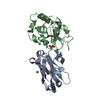
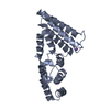
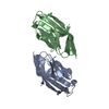
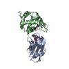
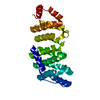
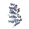
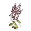
| Title | Crystal structure of restriction endonuclease HinP1I | ||||||
 Components Components | R.HinP1I restriction endonuclease | ||||||
 Keywords Keywords | HYDROLASE / Restriction endonuclease / dimerizaton | ||||||
| Function / homology | Trna Endonuclease; Chain: A, domain 1 - #40 / Restriction endonuclease, type II, HinP1I / R.HinP1I restriction endonuclease / Trna Endonuclease; Chain: A, domain 1 / endonuclease activity / 3-Layer(aba) Sandwich / metal ion binding / Alpha Beta / R.HinP1I restriction endonuclease Function and homology information Function and homology information | ||||||
| Biological species |  Haemophilus influenzae (bacteria) Haemophilus influenzae (bacteria) | ||||||
| Method |  X-RAY DIFFRACTION / SYNCHROTRON / MIR / Resolution: 2.65 Å X-RAY DIFFRACTION / SYNCHROTRON / MIR / Resolution: 2.65 Å | ||||||
 Authors Authors | Yang, Z. / Horton, J.R. / Maunus, R. / Wilson, G.G. / Roberts, R.J. / Cheng, X. | ||||||
 Citation Citation | Journal: Nucleic Acids Res. / Year: 2005 Title: Structure of HinP1I endonuclease reveals a striking similarity to the monomeric restriction enzyme MspI Authors: Yang, Z. / Horton, J.R. / Maunus, R. / Wilson, G.G. / Roberts, R.J. / Cheng, X. | ||||||
| History |
|
- Structure visualization
| Structure viewer | Molecule: MolmilJmol/JSmol |
|---|
- Downloads & links
-Download
| PDBx/mmCIF format | 1ynm.cif.gz | 62.4 KB | Display | PDBx/mmCIF format |
|---|---|---|---|---|
| PDB format | pdb1ynm.ent.gz | 46.6 KB | Display | PDB format |
| PDBx/mmJSON format | 1ynm.json.gz | Tree view | PDBx/mmJSON format | |
| Others |  Other downloads Other downloads |
-Validation report
| Arichive directory | https://data.pdbj.org/pub/pdb/validation_reports/yn/1ynmftp://data.pdbj.org/pub/pdb/validation_reports/yn/1ynm | HTTPS FTP |
|---|
-Related structure data
| Similar structure data |
|---|


-Links
 PDBj
PDBj- Assembly
| Deposited unit | 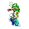
| ||||||||
|---|---|---|---|---|---|---|---|---|---|
| 1 |
| ||||||||
| Unit cell |
| ||||||||
| Details | The asymmetric unit of the crystal structure contain one biological unit. |
-Components
| #1: Protein | Mass: 28791.668 Da / Num. of mol.: 1 Source method: isolated from a genetically manipulated source Source: (gene. exp.) Haemophilus influenzae (bacteria) / Gene: hinP1IR / Plasmid: pUC19 / Production host: References: UniProt: Q5I6E6, type II site-specific deoxyribonuclease |
|---|---|
| #2: Water | ChemComp-HOH /  Mass: 18.015 Da / Num. of mol.: 100 / Source method: isolated from a natural source / Formula: H2O Mass: 18.015 Da / Num. of mol.: 100 / Source method: isolated from a natural source / Formula: H2O |
-Experimental details
-Experiment
| Experiment | Method: X-RAY DIFFRACTION / Number of used crystals: 1 |
|---|
- Sample preparation
| Crystal | Density Matthews: 4.2 Å3/Da / Density % sol: 70.3 % |
|---|---|
| Crystal grow | Temperature: 289 K / Method: vapor diffusion, hanging drop / pH: 5.5 Details: PEG 4000, sodium chloride, ammonium acetate, MES, pH 5.5, VAPOR DIFFUSION, HANGING DROP, temperature 289K |
-Data collection
| Diffraction | Mean temperature: 100 K |
|---|---|
| Diffraction source | Source: SYNCHROTRON / Site: NSLS  / Beamline: X8C / Wavelength: 1.072 Å / Beamline: X8C / Wavelength: 1.072 Å |
| Detector | Type: MARRESEARCH / Detector: CCD / Date: Mar 27, 1998 |
| Radiation | Monochromator: Si 111 CHANNEL / Protocol: SINGLE WAVELENGTH / Monochromatic (M) / Laue (L): M / Scattering type: x-ray |
| Radiation wavelength | Wavelength: 1.072 Å / Relative weight: 1 |
| Reflection | Resolution: 2.6→22 Å / Num. all: 14790 / Num. obs: 14790 / % possible obs: 99.9 % / Observed criterion σ(F): 0 / Observed criterion σ(I): 0 / Redundancy: 18.9 % / Biso Wilson estimate: 38.9 Å2 / Rmerge(I) obs: 0.075 / Net I/σ(I): 12 |
| Reflection shell | Resolution: 2.6→2.64 Å / Redundancy: 11.1 % / Rmerge(I) obs: 0.409 / Mean I/σ(I) obs: 5.1 / Num. unique all: 714 / % possible all: 99.9 |
- Processing
| Software |
| ||||||||||||||||||||||||||||||||||||||||||||||||||||||||||||
|---|---|---|---|---|---|---|---|---|---|---|---|---|---|---|---|---|---|---|---|---|---|---|---|---|---|---|---|---|---|---|---|---|---|---|---|---|---|---|---|---|---|---|---|---|---|---|---|---|---|---|---|---|---|---|---|---|---|---|---|---|---|
| Refinement | Method to determine structure: MIR / Resolution: 2.65→20 Å / Rfactor Rfree error: 0.01 / Data cutoff high absF: 1541322.66 / Data cutoff low absF: 0 / Isotropic thermal model: RESTRAINED / Cross valid method: THROUGHOUT / σ(F): 0 / Stereochemistry target values: Engh & Huber
| ||||||||||||||||||||||||||||||||||||||||||||||||||||||||||||
| Solvent computation | Solvent model: FLAT MODEL / Bsol: 40.7455 Å2 / ksol: 0.339586 e/Å3 | ||||||||||||||||||||||||||||||||||||||||||||||||||||||||||||
| Displacement parameters | Biso mean: 46.6 Å2
| ||||||||||||||||||||||||||||||||||||||||||||||||||||||||||||
| Refine analyze |
| ||||||||||||||||||||||||||||||||||||||||||||||||||||||||||||
| Refinement step | Cycle: LAST / Resolution: 2.65→20 Å
| ||||||||||||||||||||||||||||||||||||||||||||||||||||||||||||
| Refine LS restraints |
| ||||||||||||||||||||||||||||||||||||||||||||||||||||||||||||
| LS refinement shell | Resolution: 2.65→2.74 Å / Rfactor Rfree error: 0.038 / Total num. of bins used: 6
| ||||||||||||||||||||||||||||||||||||||||||||||||||||||||||||
| Xplor file |
|
