 Movie
Movie Controller


+ Open data

- Basic information
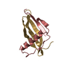
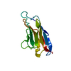
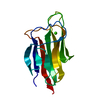
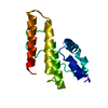
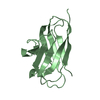
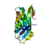
| Entry | Database: PDB / ID: 1ncn | ||||||
|---|---|---|---|---|---|---|---|
| Title | the receptor-binding domain of human B7-2 | ||||||
 Components Components | T lymphocyte activation antigen CD86 | ||||||
 Keywords Keywords | IMMUNE SYSTEM / Ig V / beta strands | ||||||
| Function / homology |  Function and homology information Function and homology informationpositive regulation of lymphotoxin A production / positive regulation of T-helper 2 cell differentiation / positive regulation of T cell receptor signaling pathway / Co-stimulation by CD28 / CD28 dependent Vav1 pathway / Co-inhibition by CTLA4 / negative regulation of T cell activation / positive regulation of immunoglobulin production / positive regulation of interleukin-4 production / Interleukin-10 signaling ...positive regulation of lymphotoxin A production / positive regulation of T-helper 2 cell differentiation / positive regulation of T cell receptor signaling pathway / Co-stimulation by CD28 / CD28 dependent Vav1 pathway / Co-inhibition by CTLA4 / negative regulation of T cell activation / positive regulation of immunoglobulin production / positive regulation of interleukin-4 production / Interleukin-10 signaling / B cell activation / CD28 dependent PI3K/Akt signaling / negative regulation of T cell receptor signaling pathway / negative regulation of T cell proliferation / coreceptor activity / T cell costimulation / centriolar satellite / positive regulation of interleukin-2 production / positive regulation of T cell proliferation / T cell activation / positive regulation of non-canonical NF-kappaB signal transduction / Constitutive Signaling by Aberrant PI3K in Cancer / PIP3 activates AKT signaling / cellular response to lipopolysaccharide / PI5P, PP2A and IER3 Regulate PI3K/AKT Signaling / virus receptor activity / signaling receptor activity / adaptive immune response / cell surface receptor signaling pathway / immune response / receptor ligand activity / external side of plasma membrane / positive regulation of cell population proliferation / positive regulation of DNA-templated transcription / cell surface / extracellular exosome / plasma membrane Similarity search - Function | ||||||
| Biological species |  Homo sapiens (human) Homo sapiens (human) | ||||||
| Method |  X-RAY DIFFRACTION / SYNCHROTRON / MAD / Resolution: 2.7 Å X-RAY DIFFRACTION / SYNCHROTRON / MAD / Resolution: 2.7 Å | ||||||
 Authors Authors | Zhang, X. / Schwartz, J.D. / Almo, S.C. / Nathenson, S.G. | ||||||
 Citation Citation | Journal: Proc.Natl.Acad.Sci.USA / Year: 2003 Title: Crystal Structure of the Receptor-Binding Domain of Human B7-2: Insights into Organization and Signaling Authors: Zhang, X. / Schwartz, J.D. / Almo, S.C. / Nathenson, S.G. #1: Journal: Protein Expr.Purif. / Year: 2002Title: Expression, Refolding, Purification, Molecular Characterization, Crystallization, and Preliminary X-ray Analysis of the Receptor Binding Domain of Human B7-2 Authors: Zhang, X. / Schwartz, J.-C.D. / Almo, S.C. / Nathenson, S.G. | ||||||
| History |
|
- Structure visualization
| Structure viewer | Molecule: MolmilJmol/JSmol |
|---|
- Downloads & links
-Download
| PDBx/mmCIF format | 1ncn.cif.gz | 54.8 KB | Display | PDBx/mmCIF format |
|---|---|---|---|---|
| PDB format | pdb1ncn.ent.gz | 40.5 KB | Display | PDB format |
| PDBx/mmJSON format | 1ncn.json.gz | Tree view | PDBx/mmJSON format | |
| Others |  Other downloads Other downloads |
-Validation report
| Arichive directory | https://data.pdbj.org/pub/pdb/validation_reports/nc/1ncnftp://data.pdbj.org/pub/pdb/validation_reports/nc/1ncn | HTTPS FTP |
|---|
-Related structure data
| Related structure data | |
|---|---|
| Similar structure data |


-Links
 PDBj
PDBj


- Assembly
| Deposited unit | 
| ||||||||
|---|---|---|---|---|---|---|---|---|---|
| 1 | 
| ||||||||
| 2 | 
| ||||||||
| Unit cell |
|
-Components
| #1: Antibody | Mass: 12849.631 Da / Num. of mol.: 2 / Fragment: Ig V-type domain Source method: isolated from a genetically manipulated source Source: (gene. exp.) Homo sapiens (human) / Gene: B7-2 / Plasmid: pET3a / Production host:  Has protein modification | Y | |
|---|
-Experimental details
-Experiment
| Experiment | Method: X-RAY DIFFRACTION / Number of used crystals: 1 |
|---|
- Sample preparation
| Crystal | Density Matthews: 2.07 Å3/Da / Density % sol: 40.18 % | ||||||||||||||||||||||||||||
|---|---|---|---|---|---|---|---|---|---|---|---|---|---|---|---|---|---|---|---|---|---|---|---|---|---|---|---|---|---|
| Crystal grow | Temperature: 277 K / Method: vapor diffusion, hanging drop / pH: 8.5 Details: PEG4000, NaAC, Tris-HCl, pH 8.5, VAPOR DIFFUSION, HANGING DROP, temperature 277K | ||||||||||||||||||||||||||||
| Crystal grow | *PLUS Temperature: 4 ℃Details: Zhang, X., (2002) PROTEIN EXPRESSION PURIF., 25, 105. | ||||||||||||||||||||||||||||
| Components of the solutions | *PLUS
|
-Data collection
| Diffraction | Mean temperature: 100 K | ||||||||||||
|---|---|---|---|---|---|---|---|---|---|---|---|---|---|
| Diffraction source | Source: SYNCHROTRON / Site: NSLS  / Beamline: X9B / Wavelength: 0.96110, 0.98000, 0.98019 / Beamline: X9B / Wavelength: 0.96110, 0.98000, 0.98019 | ||||||||||||
| Detector | Type: ADSC QUANTUM 4 / Detector: CCD / Date: Jan 1, 1999 | ||||||||||||
| Radiation | Protocol: MAD / Monochromatic (M) / Laue (L): M / Scattering type: x-ray | ||||||||||||
| Radiation wavelength |
| ||||||||||||
| Reflection | Resolution: 2.7→19.83 Å / Num. all: 5843 / Num. obs: 5843 / % possible obs: 95.5 % / Observed criterion σ(F): 0 / Observed criterion σ(I): 0 / Biso Wilson estimate: 31.2 Å2 / Limit h max: 20 / Limit h min: 0 / Limit k max: 23 / Limit k min: 0 / Limit l max: 21 / Limit l min: 0 / Observed criterion F max: 1489970.8 / Observed criterion F min: 41.08 | ||||||||||||
| Reflection shell | Resolution: 2.7→2.8 Å / % possible all: 90.8 | ||||||||||||
| Reflection | *PLUS Highest resolution: 2.7 Å / Lowest resolution: 30 Å / Num. obs: 11122 / % possible obs: 99.8 % / Num. measured all: 44045 / Rmerge(I) obs: 0.095 | ||||||||||||
| Reflection shell | *PLUS % possible obs: 100 % / Num. unique obs: 1110 / Rmerge(I) obs: 0.319 / Mean I/σ(I) obs: 3.7 |
- Processing
| Software |
| ||||||||||||||||||||||||||||||||||||||||||||||||||||||||||||||||||||||||||||||||||||||||||||||||||||||||||||||
|---|---|---|---|---|---|---|---|---|---|---|---|---|---|---|---|---|---|---|---|---|---|---|---|---|---|---|---|---|---|---|---|---|---|---|---|---|---|---|---|---|---|---|---|---|---|---|---|---|---|---|---|---|---|---|---|---|---|---|---|---|---|---|---|---|---|---|---|---|---|---|---|---|---|---|---|---|---|---|---|---|---|---|---|---|---|---|---|---|---|---|---|---|---|---|---|---|---|---|---|---|---|---|---|---|---|---|---|---|---|---|---|
| Refinement | Method to determine structure: MAD / Resolution: 2.7→19.83 Å / Rfactor Rfree error: 0.011 / Occupancy max: 1 / Occupancy min: 1 / Cross valid method: THROUGHOUT / σ(F): 2 / Stereochemistry target values: Engh & Huber
| ||||||||||||||||||||||||||||||||||||||||||||||||||||||||||||||||||||||||||||||||||||||||||||||||||||||||||||||
| Solvent computation | Solvent model: CNS bulk solvent model used / Bsol: 18.2567 Å2 / ksol: 0.37304 e/Å3 | ||||||||||||||||||||||||||||||||||||||||||||||||||||||||||||||||||||||||||||||||||||||||||||||||||||||||||||||
| Displacement parameters | Biso max: 49.42 Å2 / Biso mean: 21.94 Å2 / Biso min: 3.2 Å2
| ||||||||||||||||||||||||||||||||||||||||||||||||||||||||||||||||||||||||||||||||||||||||||||||||||||||||||||||
| Refine Biso | Class: polymer / Treatment: isotropic | ||||||||||||||||||||||||||||||||||||||||||||||||||||||||||||||||||||||||||||||||||||||||||||||||||||||||||||||
| Refine analyze |
| ||||||||||||||||||||||||||||||||||||||||||||||||||||||||||||||||||||||||||||||||||||||||||||||||||||||||||||||
| Refinement step | Cycle: LAST / Resolution: 2.7→19.83 Å
| ||||||||||||||||||||||||||||||||||||||||||||||||||||||||||||||||||||||||||||||||||||||||||||||||||||||||||||||
| Refine LS restraints |
| ||||||||||||||||||||||||||||||||||||||||||||||||||||||||||||||||||||||||||||||||||||||||||||||||||||||||||||||
| LS refinement shell | Refine-ID: X-RAY DIFFRACTION / Total num. of bins used: 10
| ||||||||||||||||||||||||||||||||||||||||||||||||||||||||||||||||||||||||||||||||||||||||||||||||||||||||||||||
| Xplor file |
| ||||||||||||||||||||||||||||||||||||||||||||||||||||||||||||||||||||||||||||||||||||||||||||||||||||||||||||||
| Software | *PLUS Name: CNS / Classification: refinement | ||||||||||||||||||||||||||||||||||||||||||||||||||||||||||||||||||||||||||||||||||||||||||||||||||||||||||||||
| Refinement | *PLUS Highest resolution: 2.7 Å / Lowest resolution: 20 Å | ||||||||||||||||||||||||||||||||||||||||||||||||||||||||||||||||||||||||||||||||||||||||||||||||||||||||||||||
| Solvent computation | *PLUS | ||||||||||||||||||||||||||||||||||||||||||||||||||||||||||||||||||||||||||||||||||||||||||||||||||||||||||||||
| Displacement parameters | *PLUS | ||||||||||||||||||||||||||||||||||||||||||||||||||||||||||||||||||||||||||||||||||||||||||||||||||||||||||||||
| Refine LS restraints | *PLUS
| ||||||||||||||||||||||||||||||||||||||||||||||||||||||||||||||||||||||||||||||||||||||||||||||||||||||||||||||
| LS refinement shell | *PLUS Highest resolution: 2.7 Å / Lowest resolution: 2.8 Å / Rfactor Rfree: 0.327 |
