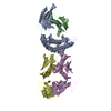
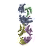
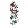
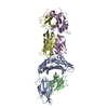
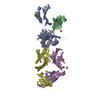
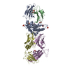
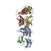
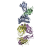
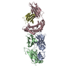
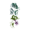
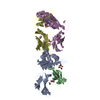
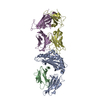
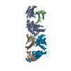
- PDB-2esv: Structure of the HLA-E-VMAPRTLIL/KK50.4 TCR complex -
+
Open data
ID or keywords:
Loading...
-
Basic information
Entry
Database: PDB / ID: 2esv
Title
Structure of the HLA-E-VMAPRTLIL/KK50.4 TCR complex
Components
(KK50.4 T cell receptor ...) x 2
Beta-2-microglobulin
HLA class I histocompatibility antigen, alpha chain E
VMAPRTLIL peptide from CMV gpUL40
Keywords
IMMUNE SYSTEM / T cell receptor / TCR / HLA-E / CMV / pMHC-TCR complex
Function / homology
Function and homology information
symbiont-mediated suppression of host natural killer cell activation / positive regulation of antibody-dependent cellular cytotoxicity / positive regulation of TRAIL production / positive regulation of natural killer cell mediated immunity / positive regulation of natural killer cell cytokine production / natural killer cell tolerance induction / regulation of natural killer cell mediated immunity / negative regulation of natural killer cell activation / antigen processing and presentation of exogenous peptide antigen via MHC class Ib / positive regulation of natural killer cell activation ...symbiont-mediated suppression of host natural killer cell activation / positive regulation of antibody-dependent cellular cytotoxicity / positive regulation of TRAIL production / positive regulation of natural killer cell mediated immunity / positive regulation of natural killer cell cytokine production / natural killer cell tolerance induction / regulation of natural killer cell mediated immunity / negative regulation of natural killer cell activation / antigen processing and presentation of exogenous peptide antigen via MHC class Ib / positive regulation of natural killer cell activation / MHC class Ib protein complex / natural killer cell lectin-like receptor binding / positive regulation of interleukin-13 production / positive regulation of natural killer cell mediated cytotoxicity / negative regulation of natural killer cell mediated cytotoxicity / alpha-beta T cell receptor complex / positive regulation of CD8-positive, alpha-beta T cell activation / CD8-positive, alpha-beta T cell activation / positive regulation of CD8-positive, alpha-beta T cell proliferation / positive regulation of immunoglobulin production / Translocation of ZAP-70 to Immunological synapse / Phosphorylation of CD3 and TCR zeta chains / positive regulation of interleukin-4 production / protection from natural killer cell mediated cytotoxicity / alpha-beta T cell activation / positive regulation of natural killer cell proliferation / Generation of second messenger molecules / beta-2-microglobulin binding / Co-inhibition by PD-1 / MHC class I protein binding / antigen processing and presentation of endogenous peptide antigen via MHC class Ib / antigen processing and presentation of endogenous peptide antigen via MHC class I via ER pathway, TAP-independent / negative regulation of T cell proliferation / T cell receptor binding / early endosome lumen / Nef mediated downregulation of MHC class I complex cell surface expression / DAP12 interactions / Endosomal/Vacuolar pathway / T cell mediated cytotoxicity / response to bacterium / Antigen Presentation: Folding, assembly and peptide loading of class I MHC / lumenal side of endoplasmic reticulum membrane / regulation of iron ion transport / cellular response to iron(III) ion / negative regulation of iron ion transport / negative regulation of forebrain neuron differentiation / antigen processing and presentation of exogenous protein antigen via MHC class Ib, TAP-dependent / peptide antigen assembly with MHC class I protein complex / ER to Golgi transport vesicle membrane / regulation of erythrocyte differentiation / response to molecule of bacterial origin / HFE-transferrin receptor complex / MHC class I peptide loading complex / transferrin transport / cellular response to iron ion / negative regulation of receptor-mediated endocytosis / positive regulation of T cell cytokine production / antigen processing and presentation of endogenous peptide antigen via MHC class I / MHC class I protein complex / peptide antigen assembly with MHC class II protein complex / negative regulation of neurogenesis / MHC class II protein complex / cellular response to nicotine / positive regulation of receptor-mediated endocytosis / multicellular organismal-level iron ion homeostasis / positive regulation of T cell mediated cytotoxicity / specific granule lumen / antigen processing and presentation of exogenous peptide antigen via MHC class II / positive regulation of immune response / peptide antigen binding / phagocytic vesicle membrane / recycling endosome membrane / positive regulation of T cell activation / negative regulation of epithelial cell proliferation / Interferon gamma signaling / Immunoregulatory interactions between a Lymphoid and a non-Lymphoid cell / positive regulation of tumor necrosis factor production / Interferon alpha/beta signaling / sensory perception of smell / Modulation by Mtb of host immune system / positive regulation of cellular senescence / tertiary granule lumen / MHC class II protein complex binding / T cell differentiation in thymus / DAP12 signaling / late endosome membrane / Downstream TCR signaling / T cell receptor signaling pathway / negative regulation of neuron projection development / antibacterial humoral response / protein refolding / ER-Phagosome pathway / early endosome membrane / amyloid fibril formation / protein homotetramerization / intracellular iron ion homeostasis / adaptive immune response / learning or memory / defense response to Gram-positive bacterium / immune response Similarity search - Function
Herpesvirus UL40 / Glycoprotein of human cytomegalovirus HHV-5 / T-cell receptor alpha chain, constant domain / Domain of unknown function (DUF1968) / : / MHC class I, alpha chain, C-terminal / MHC_I C-terminus / MHC class I-like antigen recognition-like / Murine Class I Major Histocompatibility Complex, H2-DB; Chain A, domain 1 / MHC class I alpha chain, alpha1 alpha2 domains ...Herpesvirus UL40 / Glycoprotein of human cytomegalovirus HHV-5 / T-cell receptor alpha chain, constant domain / Domain of unknown function (DUF1968) / : / MHC class I, alpha chain, C-terminal / MHC_I C-terminus / MHC class I-like antigen recognition-like / Murine Class I Major Histocompatibility Complex, H2-DB; Chain A, domain 1 / MHC class I alpha chain, alpha1 alpha2 domains / Class I Histocompatibility antigen, domains alpha 1 and 2 / Beta-2-Microglobulin / : / MHC class I-like antigen recognition-like / MHC class I-like antigen recognition-like superfamily / MHC classes I/II-like antigen recognition protein / : / Immunoglobulin/major histocompatibility complex, conserved site / Immunoglobulins and major histocompatibility complex proteins signature. / Immunoglobulin C-Type / Immunoglobulin C1-set / Immunoglobulin C1-set domain / Ig-like domain profile. / Immunoglobulin-like domain / Immunoglobulin-like domain superfamily / Immunoglobulin-like fold / Immunoglobulins / Immunoglobulin-like / Sandwich / 2-Layer Sandwich / Mainly Beta / Alpha Beta Similarity search - Domain/homology
IODIDE ION / T cell receptor alpha chain constant / T cell receptor beta constant 1 / HLA class I histocompatibility antigen, alpha chain E / Protein UL40 / Beta-2-microglobulin Similarity search - Component
Biological species
Homo sapiens (human)
Method
X-RAY DIFFRACTION / SYNCHROTRON / SAD / Resolution: 2.6 Å
Protocol: SINGLE WAVELENGTH / Monochromatic (M) / Laue (L): M / Scattering type: x-ray
Radiation wavelength
Wavelength: 1 Å / Relative weight: 1
Reflection
Highest resolution: 2.6 Å / Num. obs: 67491
Reflection shell
Highest resolution: 2.6 Å / Rmerge(I) obs: 0.104 / Mean I/σ(I) obs: 11.2 / Num. unique all: 29161
-
Processing
Software
Name
Version
Classification
NB
REFMAC
5.2.0005
refinement
PDB_EXTRACT
1.7
dataextraction
HKL-2000
datareduction
SCALEPACK
datascaling
AMoRE
phasing
Refinement
Method to determine structure: SAD / Resolution: 2.6→36.4 Å / Cor.coef. Fo:Fc: 0.926 / Cor.coef. Fo:Fc free: 0.859 / SU B: 25.202 / SU ML: 0.279 / Cross valid method: THROUGHOUT / σ(F): 0 / ESU R: 2.046 / ESU R Free: 0.391 / Stereochemistry target values: MAXIMUM LIKELIHOOD / Details: HYDROGENS HAVE BEEN ADDED IN THE RIDING POSITIONS
Rfactor
Num. reflection
% reflection
Selection details
Rfree
0.292
1467
5 %
RANDOM
Rwork
0.214
-
-
-
all
0.218
-
-
-
obs
0.218
29119
93.74 %
-
Solvent computation
Ion probe radii: 0.8 Å / Shrinkage radii: 0.8 Å / VDW probe radii: 1.2 Å / Solvent model: MASK
Displacement parameters
Biso mean: 29.311 Å2
Baniso -1
Baniso -2
Baniso -3
1-
-0.07 Å2
0 Å2
-0.83 Å2
2-
-
-0.05 Å2
0 Å2
3-
-
-
-0.56 Å2
Refine analyze
Luzzati coordinate error obs: 0.46 Å / Luzzati sigma a obs: 0.56 Å
Refinement step
Cycle: LAST / Resolution: 2.6→36.4 Å
Protein
Nucleic acid
Ligand
Solvent
Total
Num. atoms
6587
0
5
185
6777
Refine LS restraints
Refine-ID
Type
Dev ideal
Dev ideal target
Number
X-RAY DIFFRACTION
r_bond_refined_d
0.006
0.021
6768
X-RAY DIFFRACTION
r_angle_refined_deg
1.016
1.936
9202
X-RAY DIFFRACTION
r_dihedral_angle_1_deg
5.561
5
810
X-RAY DIFFRACTION
r_dihedral_angle_2_deg
35.493
23.902
346
X-RAY DIFFRACTION
r_dihedral_angle_3_deg
17.518
15
1105
X-RAY DIFFRACTION
r_dihedral_angle_4_deg
17.443
15
48
X-RAY DIFFRACTION
r_chiral_restr
0.07
0.2
975
X-RAY DIFFRACTION
r_gen_planes_refined
0.003
0.02
5280
X-RAY DIFFRACTION
r_nbd_refined
0.184
0.2
2823
X-RAY DIFFRACTION
r_nbtor_refined
0.3
0.2
4463
X-RAY DIFFRACTION
r_xyhbond_nbd_refined
0.124
0.2
317
X-RAY DIFFRACTION
r_symmetry_vdw_refined
0.174
0.2
51
X-RAY DIFFRACTION
r_symmetry_hbond_refined
0.154
0.2
9
X-RAY DIFFRACTION
r_mcbond_it
0.87
3
4105
X-RAY DIFFRACTION
r_mcangle_it
1.654
5
6625
X-RAY DIFFRACTION
r_scbond_it
2.458
7
2955
X-RAY DIFFRACTION
r_scangle_it
3.965
10
2577
LS refinement shell
Resolution: 2.6→2.668 Å / Total num. of bins used: 20
Rfactor
Num. reflection
% reflection
Rfree
0.454
102
-
Rwork
0.339
1850
-
all
-
1952
-
obs
-
-
87.18 %
+
About Yorodumi
-
News
-
Feb 9, 2022. New format data for meta-information of EMDB entries
New format data for meta-information of EMDB entries
Version 3 of the EMDB header file is now the official format.
The previous official version 1.9 will be removed from the archive.
In the structure databanks used in Yorodumi, some data are registered as the other names, "COVID-19 virus" and "2019-nCoV". Here are the details of the virus and the list of structure data.
Jan 31, 2019. EMDB accession codes are about to change! (news from PDBe EMDB page)
EMDB accession codes are about to change! (news from PDBe EMDB page)
The allocation of 4 digits for EMDB accession codes will soon come to an end. Whilst these codes will remain in use, new EMDB accession codes will include an additional digit and will expand incrementally as the available range of codes is exhausted. The current 4-digit format prefixed with “EMD-” (i.e. EMD-XXXX) will advance to a 5-digit format (i.e. EMD-XXXXX), and so on. It is currently estimated that the 4-digit codes will be depleted around Spring 2019, at which point the 5-digit format will come into force.
The EM Navigator/Yorodumi systems omit the EMD- prefix.
Related info.:Q: What is EMD? / ID/Accession-code notation in Yorodumi/EM Navigator
Yorodumi is a browser for structure data from EMDB, PDB, SASBDB, etc.
This page is also the successor to EM Navigator detail page, and also detail information page/front-end page for Omokage search.
The word "yorodu" (or yorozu) is an old Japanese word meaning "ten thousand". "mi" (miru) is to see.
Related info.:EMDB / PDB / SASBDB / Comparison of 3 databanks / Yorodumi Search / Aug 31, 2016. New EM Navigator & Yorodumi / Yorodumi Papers / Jmol/JSmol / Function and homology information / Changes in new EM Navigator and Yorodumi
 Movie
Movie Controller


 Open data
 Basic information
 Components
 Keywords
 Function and homology information
 Homo sapiens (human)
 X-RAY DIFFRACTION /
  Authors
 Citation
 Structure visualization
 Downloads & links
 Other downloads
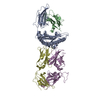


 PDBj
PDBj


 Assembly


 Mass: 126.904 Da / Num. of mol.: 5 / Source method: obtained synthetically / Formula: I
 Sample preparation
 / Beamline: 14-BM-C / Wavelength: 1 Å
 Processing