 Movie
Movie Controller

[English] 日本語
 Yorodumi
- PDB-2d74: Crystal structure of translation initiation factor aIF2betagamma ... -
+ Open data

- Basic information
| Entry | Database: PDB / ID: 2d74 | ||||||
|---|---|---|---|---|---|---|---|
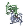
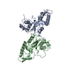
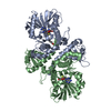
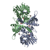
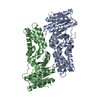
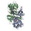
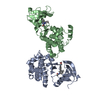
| Title | Crystal structure of translation initiation factor aIF2betagamma heterodimer | ||||||
 Components Components |
| ||||||
 Keywords Keywords | TRANSLATION / PROTEIN COMPLEX | ||||||
| Function / homology |  Function and homology information Function and homology informationformation of translation preinitiation complex / protein-synthesizing GTPase / translation elongation factor activity / translation initiation factor activity / tRNA binding / GTPase activity / GTP binding / metal ion binding / cytosol Similarity search - Function | ||||||
| Biological species |   Pyrococcus furiosus (archaea) Pyrococcus furiosus (archaea) | ||||||
| Method |  X-RAY DIFFRACTION / SYNCHROTRON / MOLECULAR REPLACEMENT / Resolution: 2.8 Å X-RAY DIFFRACTION / SYNCHROTRON / MOLECULAR REPLACEMENT / Resolution: 2.8 Å | ||||||
 Authors Authors | Sokabe, M. / Yao, M. / Sakai, N. / Toya, S. / Tanaka, I. | ||||||
 Citation Citation | Journal: Proc.Natl.Acad.Sci.USA / Year: 2006 Title: Structure of archaeal translational initiation factor 2 betagamma-GDP reveals significant conformational change of the beta-subunit and switch 1 region. Authors: Sokabe, M. / Yao, M. / Sakai, N. / Toya, S. / Tanaka, I. | ||||||
| History |
|
- Structure visualization
| Structure viewer | Molecule: MolmilJmol/JSmol |
|---|
- Downloads & links
-Download
| PDBx/mmCIF format | 2d74.cif.gz | 122.2 KB | Display | PDBx/mmCIF format |
|---|---|---|---|---|
| PDB format | pdb2d74.ent.gz | 94.3 KB | Display | PDB format |
| PDBx/mmJSON format | 2d74.json.gz | Tree view | PDBx/mmJSON format | |
| Others |  Other downloads Other downloads |
-Validation report
| Arichive directory | https://data.pdbj.org/pub/pdb/validation_reports/d7/2d74ftp://data.pdbj.org/pub/pdb/validation_reports/d7/2d74 | HTTPS FTP |
|---|
-Related structure data
| Related structure data |  2dcuC 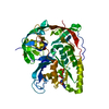 1kk1S S: Starting model for refinement C: citing same article ( |
|---|---|
| Similar structure data |


-Links
 PDBj
PDBj


- Assembly
| Deposited unit | 
| ||||||||
|---|---|---|---|---|---|---|---|---|---|
| 1 |
| ||||||||
| Unit cell |
|
-Components
| #1: Protein | Mass: 46229.484 Da / Num. of mol.: 1 / Mutation: G236D Source method: isolated from a genetically manipulated source Source: (gene. exp.) Pyrococcus furiosus (archaea) / Strain: DSM 3638 / Gene: PF1717 / Plasmid: pET26b / Production host:  | ||||
|---|---|---|---|---|---|
| #2: Protein | Mass: 17332.055 Da / Num. of mol.: 1 Source method: isolated from a genetically manipulated source Source: (gene. exp.) Pyrococcus furiosus (archaea) / Strain: DSM 3638 / Gene: PF0481 / Plasmid: pET26b / Production host: | ||||
| #3: Chemical |   Mass: 65.409 Da / Num. of mol.: 2 / Source method: obtained synthetically / Formula: Zn Mass: 65.409 Da / Num. of mol.: 2 / Source method: obtained synthetically / Formula: Zn#4: Water | ChemComp-HOH / |  Mass: 18.015 Da / Num. of mol.: 180 / Source method: isolated from a natural source / Formula: H2O Mass: 18.015 Da / Num. of mol.: 180 / Source method: isolated from a natural source / Formula: H2OHas protein modification | Y | |
-Experimental details
-Experiment
| Experiment | Method: X-RAY DIFFRACTION / Number of used crystals: 1 |
|---|
- Sample preparation
| Crystal | Density Matthews: 2.01 Å3/Da / Density % sol: 38.87 % |
|---|---|
| Crystal grow | Temperature: 293 K / Method: vapor diffusion, hanging drop / pH: 8.5 Details: 30% PEG 8000, 0.1M tris-HCl, 0.2M magnesium chloride, pH 8.5, VAPOR DIFFUSION, HANGING DROP, temperature 293K |
-Data collection
| Diffraction | Mean temperature: 100 K |
|---|---|
| Diffraction source | Source: SYNCHROTRON / Site: SPring-8  / Beamline: BL41XU / Wavelength: 0.9 Å / Beamline: BL41XU / Wavelength: 0.9 Å |
| Detector | Type: ADSC QUANTUM 315 / Detector: CCD / Date: Nov 15, 2004 |
| Radiation | Protocol: SINGLE WAVELENGTH / Monochromatic (M) / Laue (L): M / Scattering type: x-ray |
| Radiation wavelength | Wavelength: 0.9 Å / Relative weight: 1 |
| Reflection | Resolution: 2.8→50 Å / Num. all: 13070 / Num. obs: 12894 / % possible obs: 98.3 % / Observed criterion σ(F): 0 / Observed criterion σ(I): -0.2 / Redundancy: 5.3 % / Biso Wilson estimate: 62.02 Å2 / Rmerge(I) obs: 0.102 / Net I/σ(I): 16.7 |
| Reflection shell | Resolution: 2.8→2.9 Å / Redundancy: 3.2 % / Rmerge(I) obs: 0.314 / Mean I/σ(I) obs: 2 / Num. unique all: 1166 / % possible all: 91.1 |
- Processing
| Software |
| |||||||||||||||||||||||||
|---|---|---|---|---|---|---|---|---|---|---|---|---|---|---|---|---|---|---|---|---|---|---|---|---|---|---|
| Refinement | Method to determine structure: MOLECULAR REPLACEMENT Starting model: PDB ENTRY 1KK1 Resolution: 2.8→20 Å / Isotropic thermal model: Isotropic / Cross valid method: THROUGHOUT / σ(F): 0 / Stereochemistry target values: Engh & Huber
| |||||||||||||||||||||||||
| Displacement parameters | Biso mean: 61.94 Å2
| |||||||||||||||||||||||||
| Refine analyze |
| |||||||||||||||||||||||||
| Refinement step | Cycle: LAST / Resolution: 2.8→20 Å
| |||||||||||||||||||||||||
| Refine LS restraints |
| |||||||||||||||||||||||||
| LS refinement shell | Resolution: 2.8→2.9 Å
|
