 Movie
Movie Controller

[English] 日本語
 Yorodumi
- PDB-1kk1: Structure of the large gamma subunit of initiation factor eIF2 fr... -
+ Open data

- Basic information
| Entry | Database: PDB / ID: 1kk1 | ||||||
|---|---|---|---|---|---|---|---|
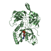
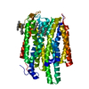
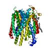
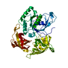
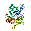
| Title | Structure of the large gamma subunit of initiation factor eIF2 from Pyrococcus abyssi-G235D mutant complexed with GDPNP-Mg2+ | ||||||
 Components Components | eIF2gamma | ||||||
 Keywords Keywords | TRANSLATION / initiation of translation | ||||||
| Function / homology |  Function and homology information Function and homology informationformation of translation preinitiation complex / protein-synthesizing GTPase / translation elongation factor activity / translation initiation factor activity / tRNA binding / GTPase activity / GTP binding / metal ion binding / cytosol Similarity search - Function | ||||||
| Biological species |   Pyrococcus abyssi (archaea) Pyrococcus abyssi (archaea) | ||||||
| Method |  X-RAY DIFFRACTION / SYNCHROTRON / rigid body minimization / Resolution: 1.8 Å X-RAY DIFFRACTION / SYNCHROTRON / rigid body minimization / Resolution: 1.8 Å | ||||||
 Authors Authors | Schmitt, E. / Blanquet, S. / Mechulam, Y. | ||||||
 Citation Citation | Journal: EMBO J. / Year: 2002 Title: The large subunit of initiation factor aIF2 is a close structural homologue of elongation factors. Authors: Schmitt, E. / Blanquet, S. / Mechulam, Y. | ||||||
| History |
|
- Structure visualization
| Structure viewer | Molecule: MolmilJmol/JSmol |
|---|
- Downloads & links
-Download
| PDBx/mmCIF format | 1kk1.cif.gz | 98.3 KB | Display | PDBx/mmCIF format |
|---|---|---|---|---|
| PDB format | pdb1kk1.ent.gz | 71.9 KB | Display | PDB format |
| PDBx/mmJSON format | 1kk1.json.gz | Tree view | PDBx/mmJSON format | |
| Others |  Other downloads Other downloads |
-Validation report
| Arichive directory | https://data.pdbj.org/pub/pdb/validation_reports/kk/1kk1ftp://data.pdbj.org/pub/pdb/validation_reports/kk/1kk1 | HTTPS FTP |
|---|
-Related structure data
| Related structure data | 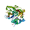 1kjzSC 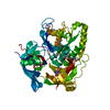 1kk0C 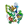 1kk2C 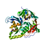 1kk3C S: Starting model for refinement C: citing same article ( |
|---|---|
| Similar structure data |


-Links
 PDBj
PDBj


- Assembly
| Deposited unit | 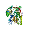
| ||||||||
|---|---|---|---|---|---|---|---|---|---|
| 1 |
| ||||||||
| Unit cell |
|
-Components
| #1: Protein | Mass: 44920.000 Da / Num. of mol.: 1 / Mutation: G235D Source method: isolated from a genetically manipulated source Source: (gene. exp.) Pyrococcus abyssi (archaea) / Plasmid: pET3a / Species (production host): Escherichia coli / Production host:  |
|---|---|
| #2: Chemical | ChemComp-ZN /   Mass: 65.409 Da / Num. of mol.: 1 / Source method: obtained synthetically / Formula: Zn Mass: 65.409 Da / Num. of mol.: 1 / Source method: obtained synthetically / Formula: Zn |
| #3: Chemical | ChemComp-MG /   Mass: 24.305 Da / Num. of mol.: 1 / Source method: obtained synthetically / Formula: Mg Mass: 24.305 Da / Num. of mol.: 1 / Source method: obtained synthetically / Formula: Mg |
| #4: Chemical | ChemComp-GNP /   Mass: 522.196 Da / Num. of mol.: 1 / Source method: obtained synthetically / Formula: C10H17N6O13P3 Mass: 522.196 Da / Num. of mol.: 1 / Source method: obtained synthetically / Formula: C10H17N6O13P3Comment: GppNHp, GMPPNP, energy-carrying molecule analogue*YM |
| #5: Water | ChemComp-HOH /  Mass: 18.015 Da / Num. of mol.: 292 / Source method: isolated from a natural source / Formula: H2O Mass: 18.015 Da / Num. of mol.: 292 / Source method: isolated from a natural source / Formula: H2O |
-Experimental details
-Experiment
| Experiment | Method: X-RAY DIFFRACTION / Number of used crystals: 1 |
|---|
- Sample preparation
| Crystal | Density Matthews: 2.7 Å3/Da / Density % sol: 54.37 % | |||||||||||||||||||||||||||||||||||||||||||||||||
|---|---|---|---|---|---|---|---|---|---|---|---|---|---|---|---|---|---|---|---|---|---|---|---|---|---|---|---|---|---|---|---|---|---|---|---|---|---|---|---|---|---|---|---|---|---|---|---|---|---|---|
| Crystal grow | Temperature: 297 K / Method: vapor diffusion, hanging drop / pH: 8.5 Details: peg8000, peg8000, tris, pH 8.5, VAPOR DIFFUSION, HANGING DROP, temperature 297K | |||||||||||||||||||||||||||||||||||||||||||||||||
| Crystal grow | *PLUS pH: 6.7 | |||||||||||||||||||||||||||||||||||||||||||||||||
| Components of the solutions | *PLUS
|
-Data collection
| Diffraction | Mean temperature: 100 K |
|---|---|
| Diffraction source | Source: SYNCHROTRON / Site: ESRF  / Beamline: ID14-1 / Wavelength: 0.934 Å / Beamline: ID14-1 / Wavelength: 0.934 Å |
| Detector | Type: ADSC QUANTUM 4 / Detector: CCD / Date: Oct 1, 2001 |
| Radiation | Monochromator: DIAMOND / Protocol: SINGLE WAVELENGTH / Monochromatic (M) / Laue (L): M / Scattering type: x-ray |
| Radiation wavelength | Wavelength: 0.934 Å / Relative weight: 1 |
| Reflection | Resolution: 1.8→45 Å / Num. all: 44188 / Num. obs: 44154 / % possible obs: 99.9 % / Observed criterion σ(F): 1 / Observed criterion σ(I): 1 / Redundancy: 3.7 % / Biso Wilson estimate: 23.6 Å2 / Rsym value: 0.043 |
| Reflection shell | Resolution: 1.8→1.85 Å / Redundancy: 3.7 % / Num. unique all: 3255 / Rsym value: 0.304 / % possible all: 100 |
| Reflection | *PLUS Rmerge(I) obs: 0.043 |
| Reflection shell | *PLUS % possible obs: 100 % / Rmerge(I) obs: 0.304 |
- Processing
| Software |
| ||||||||||||||||||||
|---|---|---|---|---|---|---|---|---|---|---|---|---|---|---|---|---|---|---|---|---|---|
| Refinement | Method to determine structure: rigid body minimization Starting model: 1kjz Resolution: 1.8→45 Å / Cross valid method: THROUGHOUT / σ(F): 0 / Stereochemistry target values: Engh & Huber
| ||||||||||||||||||||
| Displacement parameters |
| ||||||||||||||||||||
| Refinement step | Cycle: LAST / Resolution: 1.8→45 Å
| ||||||||||||||||||||
| Refine LS restraints |
| ||||||||||||||||||||
| LS refinement shell | Resolution: 1.8→1.81 Å
| ||||||||||||||||||||
| Refinement | *PLUS % reflection Rfree: 5 % / Rfactor obs: 0.219 / Rfactor Rfree: 0.245 / Rfactor Rwork: 0.219 | ||||||||||||||||||||
| Solvent computation | *PLUS | ||||||||||||||||||||
| Displacement parameters | *PLUS | ||||||||||||||||||||
| LS refinement shell | *PLUS Rfactor Rfree: 0.3848 / Rfactor Rwork: 0.2899 |
