 Movie
Movie Controller


+ Open data

- Basic information
| Entry | Database: PDB / ID: 4ak9 | ||||||
|---|---|---|---|---|---|---|---|
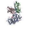
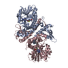
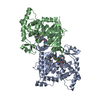
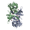
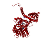
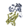
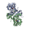
| Title | Structure of chloroplast FtsY from Physcomitrella patens | ||||||
 Components Components | CPFTSY | ||||||
 Keywords Keywords | PROTEIN TRANSPORT / CHLOROPLAST BIOGENESIS / SIMIBI GTPASE | ||||||
| Function / homology |  Function and homology information Function and homology informationSRP-dependent cotranslational protein targeting to membrane / GTP binding / membrane Similarity search - Function | ||||||
| Biological species |  PHYSCOMITRELLA PATENS (plant) PHYSCOMITRELLA PATENS (plant) | ||||||
| Method |  X-RAY DIFFRACTION / SYNCHROTRON / MOLECULAR REPLACEMENT / Resolution: 1.8 Å X-RAY DIFFRACTION / SYNCHROTRON / MOLECULAR REPLACEMENT / Resolution: 1.8 Å | ||||||
 Authors Authors | Trager, C. / Schunemann, D. / Hofmann, E. | ||||||
 Citation Citation | Journal: Plant Cell / Year: 2012 Title: Evolution from the Prokaryotic to the Higher Plant Chloroplast Signal Recognition Particle: The Signal Recognition Particle RNA is Conserved in Plastids of a Wide Range of Photosynthetic Organisms. Authors: Trager, C. / Rosenblad, M.A. / Ziehe, D. / Garcia-Petit, C. / Schrader, L. / Kock, K. / Vera Richter, C. / Klinkert, B. / Narberhaus, F. / Herrmann, C. / Hofmann, E. / Aronsson, H. / Schunemann, D. | ||||||
| History |
|
- Structure visualization
| Structure viewer | Molecule: MolmilJmol/JSmol |
|---|
- Downloads & links
-Download
| PDBx/mmCIF format | 4ak9.cif.gz | 136.5 KB | Display | PDBx/mmCIF format |
|---|---|---|---|---|
| PDB format | pdb4ak9.ent.gz | 106 KB | Display | PDB format |
| PDBx/mmJSON format | 4ak9.json.gz | Tree view | PDBx/mmJSON format | |
| Others |  Other downloads Other downloads |
-Validation report
| Arichive directory | https://data.pdbj.org/pub/pdb/validation_reports/ak/4ak9ftp://data.pdbj.org/pub/pdb/validation_reports/ak/4ak9 | HTTPS FTP |
|---|
-Related structure data
| Related structure data | 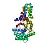 2og2S S: Starting model for refinement |
|---|---|
| Similar structure data |


-Links
 PDBj
PDBj- Assembly
| Deposited unit | 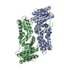
| ||||||||||||||||||||||||||||||||||||||||||||||||||||||||||
|---|---|---|---|---|---|---|---|---|---|---|---|---|---|---|---|---|---|---|---|---|---|---|---|---|---|---|---|---|---|---|---|---|---|---|---|---|---|---|---|---|---|---|---|---|---|---|---|---|---|---|---|---|---|---|---|---|---|---|---|
| 1 |
| ||||||||||||||||||||||||||||||||||||||||||||||||||||||||||
| Unit cell |
| ||||||||||||||||||||||||||||||||||||||||||||||||||||||||||
| Components on special symmetry positions |
| ||||||||||||||||||||||||||||||||||||||||||||||||||||||||||
| Noncrystallographic symmetry (NCS) | NCS domain:
NCS domain segments:
NCS oper:
|
-Components
| #1: Protein | Mass: 34560.488 Da / Num. of mol.: 2 / Fragment: RESIDUES 80-383 Source method: isolated from a genetically manipulated source Source: (gene. exp.) PHYSCOMITRELLA PATENS (plant) / Plasmid: PET-DUET-1-PP-CPFTSYD1-79 / Production host:  #2: Water | ChemComp-HOH / |  Mass: 18.015 Da / Num. of mol.: 485 / Source method: isolated from a natural source / Formula: H2O Mass: 18.015 Da / Num. of mol.: 485 / Source method: isolated from a natural source / Formula: H2OSequence details | PREDICTED TRANSIT PEPTIDE (1-56) AND 23 ADDITIONAL | |
|---|
-Experimental details
-Experiment
| Experiment | Method: X-RAY DIFFRACTION / Number of used crystals: 1 |
|---|
- Sample preparation
| Crystal | Density Matthews: 2.3 Å3/Da / Density % sol: 47 % / Description: NONE |
|---|---|
| Crystal grow | pH: 6.6 Details: 0.2M SODIUM NITRATE, 0.1M BIS-TRIS PROPANE PH6.5, 20% PEG3350, pH 6.6 |
-Data collection
| Diffraction | Mean temperature: 100 K |
|---|---|
| Diffraction source | Source: SYNCHROTRON / Site: SLS  / Beamline: X10SA / Wavelength: 1 / Beamline: X10SA / Wavelength: 1 |
| Detector | Type: DECTRIS PILATUS 6M / Detector: PIXEL / Date: May 27, 2011 / Details: MIRRORS |
| Radiation | Monochromator: SI(111) MONOCHROMATOR / Protocol: SINGLE WAVELENGTH / Monochromatic (M) / Laue (L): M / Scattering type: x-ray |
| Radiation wavelength | Wavelength: 1 Å / Relative weight: 1 |
| Reflection | Resolution: 1.8→47.7 Å / Num. obs: 58928 / % possible obs: 100 % / Observed criterion σ(I): 0 / Redundancy: 21.4 % / Biso Wilson estimate: 36 Å2 / Rmerge(I) obs: 0.14 / Net I/σ(I): 15.89 |
| Reflection shell | Resolution: 1.8→1.85 Å / Redundancy: 26.62 % / Rmerge(I) obs: 0.94 / Mean I/σ(I) obs: 4.28 / % possible all: 100 |
- Processing
| Software |
| ||||||||||||||||||||||||||||||||||||||||||||||||||||||||||||||||||||||||||||||||||||||||||||||||||||||||||||||||||||||||||||||||||||||||||||||||||||||||||
|---|---|---|---|---|---|---|---|---|---|---|---|---|---|---|---|---|---|---|---|---|---|---|---|---|---|---|---|---|---|---|---|---|---|---|---|---|---|---|---|---|---|---|---|---|---|---|---|---|---|---|---|---|---|---|---|---|---|---|---|---|---|---|---|---|---|---|---|---|---|---|---|---|---|---|---|---|---|---|---|---|---|---|---|---|---|---|---|---|---|---|---|---|---|---|---|---|---|---|---|---|---|---|---|---|---|---|---|---|---|---|---|---|---|---|---|---|---|---|---|---|---|---|---|---|---|---|---|---|---|---|---|---|---|---|---|---|---|---|---|---|---|---|---|---|---|---|---|---|---|---|---|---|---|---|---|
| Refinement | Method to determine structure: MOLECULAR REPLACEMENT Starting model: PDB ENTRY 2OG2 Resolution: 1.8→47.672 Å / SU ML: 0.19 / σ(F): 2 / Phase error: 27.12 / Stereochemistry target values: ML
| ||||||||||||||||||||||||||||||||||||||||||||||||||||||||||||||||||||||||||||||||||||||||||||||||||||||||||||||||||||||||||||||||||||||||||||||||||||||||||
| Solvent computation | Shrinkage radii: 0.98 Å / VDW probe radii: 1.2 Å / Solvent model: FLAT BULK SOLVENT MODEL / Bsol: 26.733 Å2 / ksol: 0.304 e/Å3 | ||||||||||||||||||||||||||||||||||||||||||||||||||||||||||||||||||||||||||||||||||||||||||||||||||||||||||||||||||||||||||||||||||||||||||||||||||||||||||
| Displacement parameters | Biso mean: 33 Å2
| ||||||||||||||||||||||||||||||||||||||||||||||||||||||||||||||||||||||||||||||||||||||||||||||||||||||||||||||||||||||||||||||||||||||||||||||||||||||||||
| Refinement step | Cycle: LAST / Resolution: 1.8→47.672 Å
| ||||||||||||||||||||||||||||||||||||||||||||||||||||||||||||||||||||||||||||||||||||||||||||||||||||||||||||||||||||||||||||||||||||||||||||||||||||||||||
| Refine LS restraints |
| ||||||||||||||||||||||||||||||||||||||||||||||||||||||||||||||||||||||||||||||||||||||||||||||||||||||||||||||||||||||||||||||||||||||||||||||||||||||||||
| Refine LS restraints NCS |
| ||||||||||||||||||||||||||||||||||||||||||||||||||||||||||||||||||||||||||||||||||||||||||||||||||||||||||||||||||||||||||||||||||||||||||||||||||||||||||
| LS refinement shell |
|
