 Movie
Movie Controller

[English] 日本語
 Yorodumi
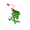
- PDB-1rqd: deoxyhypusine synthase holoenzyme in its low ionic strength, high... -
+ Open data

- Basic information
| Entry | Database: PDB / ID: 1rqd | ||||||
|---|---|---|---|---|---|---|---|
| Title | deoxyhypusine synthase holoenzyme in its low ionic strength, high pH crystal form with the inhibitor GC7 bound in the active site | ||||||
 Components Components | Deoxyhypusine synthase | ||||||
 Keywords Keywords | TRANSFERASE / Rossmann Fold / NAD cofactor / deoxyhypusine / hypusine / spermidine / GC7 | ||||||
| Function / homology |  Function and homology information Function and homology informationdeoxyhypusine synthase / Hypusine synthesis from eIF5A-lysine / deoxyhypusine synthase activity / spermidine metabolic process / spermidine catabolic process / positive regulation of T cell proliferation / protein maturation / glucose homeostasis / translation / positive regulation of cell population proliferation ...deoxyhypusine synthase / Hypusine synthesis from eIF5A-lysine / deoxyhypusine synthase activity / spermidine metabolic process / spermidine catabolic process / positive regulation of T cell proliferation / protein maturation / glucose homeostasis / translation / positive regulation of cell population proliferation / identical protein binding / cytoplasm / cytosol Similarity search - Function | ||||||
| Biological species |  Homo sapiens (human) Homo sapiens (human) | ||||||
| Method |  X-RAY DIFFRACTION / SYNCHROTRON / FOURIER SYNTHESIS / Resolution: 3 Å X-RAY DIFFRACTION / SYNCHROTRON / FOURIER SYNTHESIS / Resolution: 3 Å | ||||||
 Authors Authors | Umland, T.C. / Wolff, E.C. / Park, M.-H. / Davies, D.R. | ||||||
 Citation Citation | Journal: J.Biol.Chem. / Year: 2004 Title: A New Crystal Structure of Deoxyhypusine Synthase Reveals the Configuration of the Active Enzyme and of an Enzyme-NAD-Inhibitor Ternary Complex Authors: Umland, T.C. / Wolff, E.C. / Park, M.-H. / Davies, D.R. #1: Journal: Structure / Year: 1998Title: Crystal structure of the NAD complex of human deoxyhypusine synthase: an enzyme with a ball-and-chain mechanism for blocking the active site Authors: Liao, D.I. / Wolff, E. / Park, M.-H. / Davies, D.R. | ||||||
| History |
|
- Structure visualization
| Structure viewer | Molecule: MolmilJmol/JSmol |
|---|
- Downloads & links
-Download
| PDBx/mmCIF format | 1rqd.cif.gz | 141.9 KB | Display | PDBx/mmCIF format |
|---|---|---|---|---|
| PDB format | pdb1rqd.ent.gz | 111.6 KB | Display | PDB format |
| PDBx/mmJSON format | 1rqd.json.gz | Tree view | PDBx/mmJSON format | |
| Others |  Other downloads Other downloads |
-Validation report
| Arichive directory | https://data.pdbj.org/pub/pdb/validation_reports/rq/1rqdftp://data.pdbj.org/pub/pdb/validation_reports/rq/1rqd | HTTPS FTP |
|---|
-Related structure data
| Related structure data | 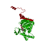 1rlzC  1rozSC C: citing same article ( S: Starting model for refinement |
|---|---|
| Similar structure data |


-Links
 PDBj
PDBj

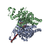
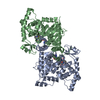
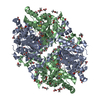
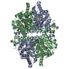
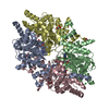
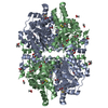
- Assembly
| Deposited unit | 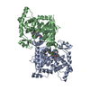
| ||||||||
|---|---|---|---|---|---|---|---|---|---|
| 1 | 
| ||||||||
| Unit cell |
| ||||||||
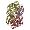
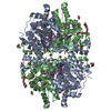
| Details | The biological assembly is a tetramer, and may be generated from the dimer contained within the asymmetric unit by the crystallographic two fold axis: x-y, -y, -z+1/3 |
-Components
| #1: Protein | Mass: 41012.434 Da / Num. of mol.: 2 Source method: isolated from a genetically manipulated source Source: (gene. exp.) Homo sapiens (human) / Gene: DHPS, DS / Plasmid: pET-11a / Species (production host): Escherichia coli / Production host:  #2: Chemical |   Mass: 663.425 Da / Num. of mol.: 2 / Source method: obtained synthetically / Formula: C21H27N7O14P2 / Comment: NAD*YM Mass: 663.425 Da / Num. of mol.: 2 / Source method: obtained synthetically / Formula: C21H27N7O14P2 / Comment: NAD*YM#3: Chemical | 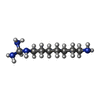  Mass: 174.287 Da / Num. of mol.: 2 / Source method: obtained synthetically / Formula: C8H22N4 Mass: 174.287 Da / Num. of mol.: 2 / Source method: obtained synthetically / Formula: C8H22N4 |
|---|
-Experimental details
-Experiment
| Experiment | Method: X-RAY DIFFRACTION / Number of used crystals: 1 |
|---|
- Sample preparation
| Crystal | Density Matthews: 3.08 Å3/Da / Density % sol: 60 % |
|---|---|
| Crystal grow | Temperature: 298 K / Method: vapor diffusion, hanging drop / pH: 8 Details: Tris-HCl, KCl, NAD, 2-methyl-2,4-pentanediol,1-guanidinium-7-aminoheptane, pH 8.0, VAPOR DIFFUSION, HANGING DROP, temperature 298K |
-Data collection
| Diffraction | Mean temperature: 95 K |
|---|---|
| Diffraction source | Source: SYNCHROTRON / Site: NSLS  / Beamline: X9B / Wavelength: 0.98 Å / Beamline: X9B / Wavelength: 0.98 Å |
| Detector | Type: ADSC QUANTUM 4 / Detector: CCD / Date: Jun 17, 1999 / Details: monochromator |
| Radiation | Monochromator: Si (111) / Protocol: SINGLE WAVELENGTH / Monochromatic (M) / Laue (L): M / Scattering type: x-ray |
| Radiation wavelength | Wavelength: 0.98 Å / Relative weight: 1 |
| Reflection | Resolution: 3→50 Å / Num. all: 20171 / Num. obs: 20171 / % possible obs: 96.3 % / Observed criterion σ(I): -3 / Redundancy: 3.4 % / Rsym value: 0.099 / Net I/σ(I): 9.1 |
| Reflection shell | Resolution: 3→3.11 Å / Redundancy: 2.8 % / Num. unique all: 1913 / Rsym value: 0.254 / % possible all: 92.5 |
- Processing
| Software |
| |||||||||||||||||||||||||
|---|---|---|---|---|---|---|---|---|---|---|---|---|---|---|---|---|---|---|---|---|---|---|---|---|---|---|
| Refinement | Method to determine structure: FOURIER SYNTHESIS Starting model: PDB ENTRY 1ROZ Resolution: 3→35 Å / Isotropic thermal model: isotropic / Cross valid method: THROUGHOUT / σ(F): 0 / σ(I): 0 / Stereochemistry target values: Engh & Huber
| |||||||||||||||||||||||||
| Displacement parameters | Biso mean: 41.6 Å2 | |||||||||||||||||||||||||
| Refine analyze |
| |||||||||||||||||||||||||
| Refinement step | Cycle: LAST / Resolution: 3→35 Å
| |||||||||||||||||||||||||
| Refine LS restraints |
|
