 Movie
Movie Controller


+ Open data

- Basic information
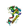
| Entry | Database: PDB / ID: 2azo | ||||||
|---|---|---|---|---|---|---|---|
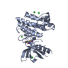
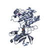
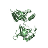
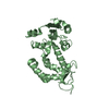
| Title | DNA MISMATCH REPAIR PROTEIN MUTH FROM E. COLI | ||||||
 Components Components | MUTH | ||||||
 Keywords Keywords | ENDONUCLEASE / MUTH / DNA REPAIR / HYDROLASE | ||||||
| Function / homology |  Function and homology information Function and homology informationT/G mismatch-specific endonuclease activity / mismatch repair complex / regulation of DNA recombination / DNA modification / mismatch repair / endonuclease activity / hydrolase activity / DNA binding / cytoplasm Similarity search - Function | ||||||
| Biological species |  | ||||||
| Method |  X-RAY DIFFRACTION / SIRAS / Resolution: 2.3 Å X-RAY DIFFRACTION / SIRAS / Resolution: 2.3 Å | ||||||
 Authors Authors | Yang, W. | ||||||
 Citation Citation | Journal: EMBO J. / Year: 1998 Title: Structural basis for MutH activation in E.coli mismatch repair and relationship of MutH to restriction endonucleases. Authors: Ban, C. / Yang, W. | ||||||
| History |
|
- Structure visualization
| Structure viewer | Molecule: MolmilJmol/JSmol |
|---|
- Downloads & links
-Download
| PDBx/mmCIF format | 2azo.cif.gz | 99.7 KB | Display | PDBx/mmCIF format |
|---|---|---|---|---|
| PDB format | pdb2azo.ent.gz | 77.1 KB | Display | PDB format |
| PDBx/mmJSON format | 2azo.json.gz | Tree view | PDBx/mmJSON format | |
| Others |  Other downloads Other downloads |
-Validation report
| Arichive directory | https://data.pdbj.org/pub/pdb/validation_reports/az/2azoftp://data.pdbj.org/pub/pdb/validation_reports/az/2azo | HTTPS FTP |
|---|
-Related structure data


-Links
 PDBj
PDBj


- Assembly
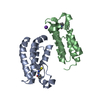
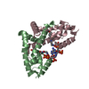
| Deposited unit | 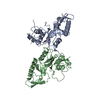
| ||||||||
|---|---|---|---|---|---|---|---|---|---|
| 1 | 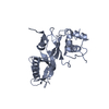
| ||||||||
| 2 | 
| ||||||||
| Unit cell |
| ||||||||
| Noncrystallographic symmetry (NCS) | NCS oper: (Code: given Matrix: (0.915447, -0.117429, 0.385006), Vector: Details | ALTHOUGH THERE ARE TWO COPIES OF MUTH IN ONE ASYMMETRIC UNITS, THE COPIES ARE STRUCTURAL DIFFERENT DUE TO CONFORMATIONAL CHANGES BETWEEN TWO SUBDOMAINS IN THE PROTEIN. | |
-Components
| #1: Protein | Mass: 25840.695 Da / Num. of mol.: 2 Source method: isolated from a genetically manipulated source Source: (gene. exp.) #2: Water | ChemComp-HOH / |  Mass: 18.015 Da / Num. of mol.: 144 / Source method: isolated from a natural source / Formula: H2O Mass: 18.015 Da / Num. of mol.: 144 / Source method: isolated from a natural source / Formula: H2O |
|---|
-Experimental details
-Experiment
| Experiment | Method: X-RAY DIFFRACTION / Number of used crystals: 1 |
|---|
- Sample preparation
| Crystal | Density Matthews: 2.35 Å3/Da / Density % sol: 48 % | |||||||||||||||||||||||||
|---|---|---|---|---|---|---|---|---|---|---|---|---|---|---|---|---|---|---|---|---|---|---|---|---|---|---|
| Crystal grow | pH: 6 Details: 100MM AMMONIUM ACETATE, 50 MM MAGNESIUM ACETATE, 1 MM DTT AND 12-16 % PEG6000, pH 6.0 | |||||||||||||||||||||||||
| Crystal grow | *PLUS Temperature: 20 ℃ / pH: 5.8 / Method: vapor diffusion, hanging drop | |||||||||||||||||||||||||
| Components of the solutions | *PLUS
|
-Data collection
| Diffraction | Mean temperature: 98 K |
|---|---|
| Diffraction source | Wavelength: 1.5418 |
| Detector | Type: RIGAKU RAXIS II / Detector: IMAGE PLATE / Date: Apr 1, 1997 / Details: MIRRORS |
| Radiation | Monochromator: NI FILTER / Monochromatic (M) / Laue (L): M / Scattering type: x-ray |
| Radiation wavelength | Wavelength: 1.5418 Å / Relative weight: 1 |
| Reflection | Resolution: 2.3→20 Å / Num. obs: 20757 / % possible obs: 95.2 % / Observed criterion σ(I): 0 / Redundancy: 2.6 % / Biso Wilson estimate: 20 Å2 / Rmerge(I) obs: 0.079 / Rsym value: 0.079 / Net I/σ(I): 10 |
| Reflection shell | Resolution: 2.28→2.37 Å / Redundancy: 2.1 % / Rmerge(I) obs: 0.369 / Mean I/σ(I) obs: 2 / Rsym value: 0.369 / % possible all: 83.8 |
| Reflection shell | *PLUS % possible obs: 83.8 % |
- Processing
| Software |
| ||||||||||||||||||||||||||||||||||||||||||||||||||||||||||||||||||||||||||||||||
|---|---|---|---|---|---|---|---|---|---|---|---|---|---|---|---|---|---|---|---|---|---|---|---|---|---|---|---|---|---|---|---|---|---|---|---|---|---|---|---|---|---|---|---|---|---|---|---|---|---|---|---|---|---|---|---|---|---|---|---|---|---|---|---|---|---|---|---|---|---|---|---|---|---|---|---|---|---|---|---|---|---|
| Refinement | Method to determine structure: SIRAS / Resolution: 2.3→20 Å / Rfactor Rfree error: 0.007 / Data cutoff high absF: 10000000 / Data cutoff low absF: 0.001 / Isotropic thermal model: RESTRAINED / Cross valid method: THROUGHOUT / σ(F): 1 / Details: BULK SOLVENT MODEL USED
| ||||||||||||||||||||||||||||||||||||||||||||||||||||||||||||||||||||||||||||||||
| Displacement parameters | Biso mean: 29.9 Å2 | ||||||||||||||||||||||||||||||||||||||||||||||||||||||||||||||||||||||||||||||||
| Refine analyze |
| ||||||||||||||||||||||||||||||||||||||||||||||||||||||||||||||||||||||||||||||||
| Refinement step | Cycle: LAST / Resolution: 2.3→20 Å
| ||||||||||||||||||||||||||||||||||||||||||||||||||||||||||||||||||||||||||||||||
| Refine LS restraints |
| ||||||||||||||||||||||||||||||||||||||||||||||||||||||||||||||||||||||||||||||||
| LS refinement shell | Resolution: 2.3→2.44 Å / Rfactor Rfree error: 0.021 / Total num. of bins used: 6
| ||||||||||||||||||||||||||||||||||||||||||||||||||||||||||||||||||||||||||||||||
| Xplor file |
| ||||||||||||||||||||||||||||||||||||||||||||||||||||||||||||||||||||||||||||||||
| Software | *PLUS Name: X-PLOR / Version: 3.851 / Classification: refinement | ||||||||||||||||||||||||||||||||||||||||||||||||||||||||||||||||||||||||||||||||
| Refinement | *PLUS Rfactor obs: 0.225 / Rfactor Rfree: 0.289 | ||||||||||||||||||||||||||||||||||||||||||||||||||||||||||||||||||||||||||||||||
| Solvent computation | *PLUS | ||||||||||||||||||||||||||||||||||||||||||||||||||||||||||||||||||||||||||||||||
| Displacement parameters | *PLUS | ||||||||||||||||||||||||||||||||||||||||||||||||||||||||||||||||||||||||||||||||
| Refine LS restraints | *PLUS
| ||||||||||||||||||||||||||||||||||||||||||||||||||||||||||||||||||||||||||||||||
| LS refinement shell | *PLUS Rfactor obs: 0.3 |
