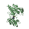
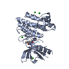
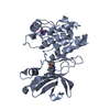
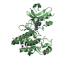
[English] 日本語
Yorodumi
- PDB-6nyh: Structure of human RIPK1 kinase domain in complex with GNE684 -

+
Open data


ID or keywords:

Loading...

-
Basic information

Entry
Database: PDB / ID: 6nyh
TitleStructure of human RIPK1 kinase domain in complex with GNE684
ComponentsReceptor-interacting serine/threonine-protein kinase 1
KeywordsIMMUNE SYSTEM / RIP1 / kinase / RIP / RIP1K RIPK1 / Transferase-Transferase inhibitor complex
Function / homology
Function and homology information


regulation of ATP:ADP antiporter activity / ripoptosome assembly / positive regulation of interleukin-6-mediated signaling pathway / positive regulation of miRNA processing / ripoptosome assembly involved in necroptotic process / death domain binding / ripoptosome / Defective RIPK1-mediated regulated necrosis / TRIF-mediated programmed cell death / TLR3-mediated TICAM1-dependent programmed cell death ...regulation of ATP:ADP antiporter activity / ripoptosome assembly / positive regulation of interleukin-6-mediated signaling pathway / positive regulation of miRNA processing / ripoptosome assembly involved in necroptotic process / death domain binding / ripoptosome / Defective RIPK1-mediated regulated necrosis / TRIF-mediated programmed cell death / TLR3-mediated TICAM1-dependent programmed cell death / Microbial modulation of RIPK1-mediated regulated necrosis / Regulation by c-FLIP / CASP8 activity is inhibited / Dimerization of procaspase-8 / programmed necrotic cell death / TNF signaling / Caspase activation via Death Receptors in the presence of ligand / T cell apoptotic process / SARS-CoV-1-mediated effects on programmed cell death / positive regulation of macrophage differentiation / necroptotic signaling pathway / JUN kinase kinase kinase activity / peptidyl-serine autophosphorylation / NF-kB activation through FADD/RIP-1 pathway mediated by caspase-8 and -10 / positive regulation of necroptotic process / RIP-mediated NFkB activation via ZBP1 / death-inducing signaling complex / negative regulation of necroptotic process / positive regulation of tumor necrosis factor-mediated signaling pathway / death receptor binding / positive regulation of extrinsic apoptotic signaling pathway / positive regulation of programmed cell death / TRP channels / positive regulation of programmed necrotic cell death / TNFR1-induced proapoptotic signaling / RIPK1-mediated regulated necrosis / positive regulation of execution phase of apoptosis / necroptotic process / negative regulation of extrinsic apoptotic signaling pathway in absence of ligand / response to tumor necrosis factor / signaling adaptor activity / extrinsic apoptotic signaling pathway / negative regulation of canonical NF-kappaB signal transduction / tumor necrosis factor-mediated signaling pathway / TICAM1, RIP1-mediated IKK complex recruitment / IKK complex recruitment mediated by RIP1 / TNFR1-induced NF-kappa-B signaling pathway / positive regulation of interleukin-8 production / negative regulation of extrinsic apoptotic signaling pathway / Regulation of TNFR1 signaling / positive regulation of JNK cascade / protein catabolic process / Regulation of necroptotic cell death / cellular response to growth factor stimulus / cellular response to hydrogen peroxide / positive regulation of inflammatory response / positive regulation of non-canonical NF-kappaB signal transduction / positive regulation of tumor necrosis factor production / positive regulation of reactive oxygen species metabolic process / positive regulation of neuron apoptotic process / Ovarian tumor domain proteases / cellular response to tumor necrosis factor / positive regulation of NF-kappaB transcription factor activity / positive regulation of canonical NF-kappaB signal transduction / response to oxidative stress / amyloid fibril formation / Potential therapeutics for SARS / protein autophosphorylation / receptor complex / non-specific serine/threonine protein kinase / endosome membrane / Ub-specific processing proteases / protein kinase activity / intracellular signal transduction / inflammatory response / positive regulation of protein phosphorylation / positive regulation of apoptotic process / protein serine kinase activity / protein serine/threonine kinase activity / apoptotic process / ubiquitin protein ligase binding / protein-containing complex binding / positive regulation of gene expression / negative regulation of apoptotic process / protein homodimerization activity / positive regulation of transcription by RNA polymerase II / protein-containing complex / mitochondrion / ATP binding / identical protein binding / plasma membrane / cytosol
Similarity search - Function
RIP1, Death domain / Death domain profile. / DEATH domain, found in proteins involved in cell death (apoptosis). / Death domain / Death domain / Death-like domain superfamily / Protein tyrosine and serine/threonine kinase / Serine-threonine/tyrosine-protein kinase, catalytic domain / Transferase(Phosphotransferase) domain 1 / Transferase(Phosphotransferase); domain 1 ...RIP1, Death domain / Death domain profile. / DEATH domain, found in proteins involved in cell death (apoptosis). / Death domain / Death domain / Death-like domain superfamily / Protein tyrosine and serine/threonine kinase / Serine-threonine/tyrosine-protein kinase, catalytic domain / Transferase(Phosphotransferase) domain 1 / Transferase(Phosphotransferase); domain 1 / Phosphorylase Kinase; domain 1 / Phosphorylase Kinase; domain 1 / Serine/threonine-protein kinase, active site / Serine/Threonine protein kinases active-site signature. / Serine/Threonine protein kinases, catalytic domain / Protein kinase domain profile. / Protein kinase domain / Protein kinase-like domain superfamily / 2-Layer Sandwich / Orthogonal Bundle / Mainly Alpha / Alpha Beta
Similarity search - Domain/homology
IODIDE ION / Chem-L8D / Receptor-interacting serine/threonine-protein kinase 1
Similarity search - Component
Biological speciesHomo sapiens (human)
MethodX-RAY DIFFRACTION / SYNCHROTRON / MOLECULAR REPLACEMENT / Resolution: 2.1 Å
AuthorsFong, R. / Lupardus, P.J.
CitationJournal: Cell Death Differ. / Year: 2020
Title: RIP1 inhibition blocks inflammatory diseases but not tumor growth or metastases.
Authors: Patel, S. / Webster, J.D. / Varfolomeev, E. / Kwon, Y.C. / Cheng, J.H. / Zhang, J. / Dugger, D.L. / Wickliffe, K.E. / Maltzman, A. / Sujatha-Bhaskar, S. / Bir Kohli, P. / Ramaswamy, S. / ...Authors: Patel, S. / Webster, J.D. / Varfolomeev, E. / Kwon, Y.C. / Cheng, J.H. / Zhang, J. / Dugger, D.L. / Wickliffe, K.E. / Maltzman, A. / Sujatha-Bhaskar, S. / Bir Kohli, P. / Ramaswamy, S. / Deshmukh, G. / Liederer, B.M. / Fong, R. / Hamilton, G. / Lupardus, P. / Caplazi, P. / Lee, W.P. / van Lookeren Campagne, M. / Johnson, A. / McKenzie, B.S. / Junttila, M.R. / Newton, K. / Vucic, D.
History
DepositionFeb 11, 2019Deposition site: RCSB / Processing site: RCSB
Revision 1.0May 29, 2019Provider: repository / Type: Initial release
Revision 1.1Jan 1, 2020Group: Database references / Category: citation / citation_author
Item: _citation.journal_volume / _citation.page_first ..._citation.journal_volume / _citation.page_first / _citation.page_last / _citation.year / _citation_author.identifier_ORCID
Revision 1.2Oct 11, 2023Group: Data collection / Database references / Refinement description
Category: chem_comp_atom / chem_comp_bond ...chem_comp_atom / chem_comp_bond / database_2 / pdbx_initial_refinement_model
Item: _database_2.pdbx_DOI / _database_2.pdbx_database_accession

-
Structure visualization

Structure viewerMolecule:
MolmilJmol/JSmol

Downloads & links

-
Assembly

Deposited unit
A: Receptor-interacting serine/threonine-protein kinase 1
B: Receptor-interacting serine/threonine-protein kinase 1
hetero molecules


Theoretical massNumber of molelcules
Total (without water)68,85210
Polymers67,2262
Non-polymers1,6268
Water3,621201
1
A: Receptor-interacting serine/threonine-protein kinase 1
hetero molecules


Theoretical massNumber of molelcules
Total (without water)34,4265
Polymers33,6131
Non-polymers8134
Water181
TypeNameSymmetry operationNumber
identity operation1_555x,y,z1
MethodPISA
2
B: Receptor-interacting serine/threonine-protein kinase 1
hetero molecules


Theoretical massNumber of molelcules
Total (without water)34,4265
Polymers33,6131
Non-polymers8134
Water181
TypeNameSymmetry operationNumber
identity operation1_555x,y,z1
MethodPISA
Unit cell
Length a, b, c (Å)46.980, 97.034, 125.238
Angle α, β, γ (deg.)90.00, 90.00, 90.00
Int Tables number19
Space group name H-MP212121

-
Components

#1: Protein Receptor-interacting serine/threonine-protein kinase 1 / Cell death protein RIP / Receptor-interacting protein 1 / RIP-1


Mass: 33612.855 Da / Num. of mol.: 2 / Mutation: C34A, C127A, C233A, C240A
Source method: isolated from a genetically manipulated source
Source: (gene. exp.) Homo sapiens (human) / Gene: RIPK1, RIP, RIP1 / Production host: 
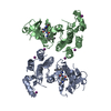
Spodoptera frugiperda (fall armyworm)
References: UniProt: Q13546, non-specific serine/threonine protein kinase
#2: Chemical ChemComp-L8D / (5S)-N-[(3S)-7-methoxy-1-methyl-2-oxo-2,3,4,5-tetrahydro-1H-pyrido[3,4-b]azepin-3-yl]-5-phenyl-6,7-dihydro-5H-pyrrolo[1,2-b][1,2,4]triazole-2-carboxamide


Mass: 432.475 Da / Num. of mol.: 2 / Source method: obtained synthetically / Formula: C23H24N6O3
#3: Chemical
ChemComp-IOD / IODIDE ION


Mass: 126.904 Da / Num. of mol.: 6 / Source method: obtained synthetically / Formula: I
#4: Water ChemComp-HOH / water


Mass: 18.015 Da / Num. of mol.: 201 / Source method: isolated from a natural source / Formula: H2O

-
Experimental details

-
Experiment

ExperimentMethod: X-RAY DIFFRACTION / Number of used crystals: 1

-
Sample preparation

CrystalDensity Matthews: 2.12 Å3/Da / Density % sol: 42.07 %
Crystal growTemperature: 277 K / Method: vapor diffusion
Details: 0.1 M Bis Tris Propane buffer (pH 6.5), 0.2 M sodium iodide, 20% PEG3350

-
Data collection

DiffractionMean temperature: 100 K / Serial crystal experiment: N
Diffraction sourceSource: SYNCHROTRON / Site: APS / Beamline: 21-ID-D / Wavelength: 1 Å
DetectorType: DECTRIS EIGER X 16M / Detector: PIXEL / Date: Aug 8, 2018
RadiationProtocol: SINGLE WAVELENGTH / Monochromatic (M) / Laue (L): M / Scattering type: x-ray
Radiation wavelengthWavelength: 1 Å / Relative weight: 1
ReflectionResolution: 2.1→48.52 Å / Num. obs: 32257 / % possible obs: 94.4 % / Redundancy: 5.7 % / Biso Wilson estimate: 30.2 Å2 / CC1/2: 0.992 / Rmerge(I) obs: 0.106 / Net I/σ(I): 8.8
Reflection shellResolution: 2.1→2.16 Å / Redundancy: 5.7 % / Rmerge(I) obs: 0.872 / Mean I/σ(I) obs: 1.6 / CC1/2: 0.723 / % possible all: 96.6

-
Processing

Software
NameVersionClassification
PHENIX1.10.1-2155_2155refinement
XDSdata reduction
Aimlessdata scaling
PHASERphasing
RefinementMethod to determine structure: MOLECULAR REPLACEMENT
Starting model: 4ITH
Resolution: 2.1→48.517 Å / SU ML: 0.29 / Cross valid method: FREE R-VALUE / σ(F): 1.34 / Phase error: 27.25
RfactorNum. reflection% reflection
Rfree0.2464 2154 6.69 %
Rwork0.2025 --
obs0.2056 32202 93.99 %
Solvent computationShrinkage radii: 0.9 Å / VDW probe radii: 1.11 Å
Refinement stepCycle: LAST / Resolution: 2.1→48.517 Å
ProteinNucleic acidLigandSolventTotal
Num. atoms4024 0 70 201 4295
Refine LS restraints
Refine-IDTypeDev idealNumber
X-RAY DIFFRACTIONf_bond_d0.0084176
X-RAY DIFFRACTIONf_angle_d0.9355639
X-RAY DIFFRACTIONf_dihedral_angle_d13.2922509
X-RAY DIFFRACTIONf_chiral_restr0.054628
X-RAY DIFFRACTIONf_plane_restr0.005706
LS refinement shell
Resolution (Å)Rfactor RfreeNum. reflection RfreeRfactor RworkNum. reflection RworkRefine-ID% reflection obs (%)
2.1-2.14890.33351380.2712029X-RAY DIFFRACTION96
2.1489-2.20260.30181250.2581879X-RAY DIFFRACTION90
2.2026-2.26220.33581050.24441618X-RAY DIFFRACTION77
2.2622-2.32870.27531570.23262064X-RAY DIFFRACTION98
2.3287-2.40390.30171380.22942065X-RAY DIFFRACTION98
2.4039-2.48980.29731490.21922052X-RAY DIFFRACTION98
2.4898-2.58950.27191700.22692066X-RAY DIFFRACTION99
2.5895-2.70730.33021240.22181901X-RAY DIFFRACTION89
2.7073-2.85010.25591580.21712065X-RAY DIFFRACTION98
2.8501-3.02860.27311440.22662062X-RAY DIFFRACTION97
3.0286-3.26240.24521380.21261935X-RAY DIFFRACTION90
3.2624-3.59060.26651430.19391981X-RAY DIFFRACTION92
3.5906-4.110.2251370.16752009X-RAY DIFFRACTION94
4.11-5.17720.18741560.16152162X-RAY DIFFRACTION98
5.1772-48.52990.21881720.21022160X-RAY DIFFRACTION95

+
About Yorodumi

-
News

-
Feb 9, 2022. New format data for meta-information of EMDB entries

New format data for meta-information of EMDB entries

  • Version 3 of the EMDB header file is now the official format.
  • The previous official version 1.9 will be removed from the archive.

Related info.:EMDB header

External links:wwPDB to switch to version 3 of the EMDB data model

-
Aug 12, 2020. Covid-19 info

Covid-19 info

URL: https://pdbjlvh1.pdbj.org/emnavi/covid19.php

New page: Covid-19 featured information page in EM Navigator.

Related info.:Covid-19 info / Mar 5, 2020. Novel coronavirus structure data

+
Mar 5, 2020. Novel coronavirus structure data

Novel coronavirus structure data

Related info.:Yorodumi Speices / Aug 12, 2020. Covid-19 info

External links:COVID-19 featured content - PDBj / Molecule of the Month (242):Coronavirus Proteases

+
Jan 31, 2019. EMDB accession codes are about to change! (news from PDBe EMDB page)

EMDB accession codes are about to change! (news from PDBe EMDB page)

  • The allocation of 4 digits for EMDB accession codes will soon come to an end. Whilst these codes will remain in use, new EMDB accession codes will include an additional digit and will expand incrementally as the available range of codes is exhausted. The current 4-digit format prefixed with “EMD-” (i.e. EMD-XXXX) will advance to a 5-digit format (i.e. EMD-XXXXX), and so on. It is currently estimated that the 4-digit codes will be depleted around Spring 2019, at which point the 5-digit format will come into force.
  • The EM Navigator/Yorodumi systems omit the EMD- prefix.

Related info.:Q: What is EMD? / ID/Accession-code notation in Yorodumi/EM Navigator

External links:EMDB Accession Codes are Changing Soon! / Contact to PDBj

+
Jul 12, 2017. Major update of PDB

Major update of PDB

  • wwPDB released updated PDB data conforming to the new PDBx/mmCIF dictionary.
  • This is a major update changing the version number from 4 to 5, and with Remediation, in which all the entries are updated.
  • In this update, many items about electron microscopy experimental information are reorganized (e.g. em_software).
  • Now, EM Navigator and Yorodumi are based on the updated data.

External links:wwPDB Remediation / Enriched Model Files Conforming to OneDep Data Standards Now Available in the PDB FTP Archive

-
Yorodumi

Thousand views of thousand structures

  • Yorodumi is a browser for structure data from EMDB, PDB, SASBDB, etc.
  • This page is also the successor to EM Navigator detail page, and also detail information page/front-end page for Omokage search.
  • The word "yorodu" (or yorozu) is an old Japanese word meaning "ten thousand". "mi" (miru) is to see.

Related info.:EMDB / PDB / SASBDB / Comparison of 3 databanks / Yorodumi Search / Aug 31, 2016. New EM Navigator & Yorodumi / Yorodumi Papers / Jmol/JSmol / Function and homology information / Changes in new EM Navigator and Yorodumi

Read more