[English] 日本語
Yorodumi
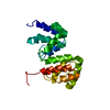
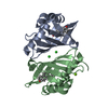
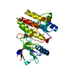
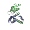
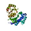
- PDB-1azo: DNA MISMATCH REPAIR PROTEIN MUTH FROM E. COLI -

+
Open data


ID or keywords:

Loading...

-
Basic information

Entry
Database: PDB / ID: 1azo
TitleDNA MISMATCH REPAIR PROTEIN MUTH FROM E. COLI
ComponentsMUTH
KeywordsENDONUCLEASE / MUTH / DNA REPAIR / HYDROLASE
Function / homology
Function and homology information


T/G mismatch-specific endonuclease activity / mismatch repair complex / regulation of DNA recombination / DNA modification / mismatch repair / endonuclease activity / DNA binding / cytoplasm
Similarity search - Function
DNA mismatch repair protein MutH / DNA mismatch repair MutH/Type II restriction enzyme Sau3AI / DNA mismatch repair enzyme MutH / DNA mismatch repair enzyme MutH / DNA mismatch repair MutH/Restriction endonuclease, type II / DNA mismatch repair MutH/Type II restriction endonuclease superfamily / ECO RV Endonuclease; Chain A / Restriction endonuclease type II-like / 3-Layer(aba) Sandwich / Alpha Beta
Similarity search - Domain/homology
DNA mismatch repair protein MutH
Similarity search - Component
Biological speciesEscherichia coli (E. coli)
MethodX-RAY DIFFRACTION / Resolution: 1.7 Å
AuthorsYang, W.
CitationJournal: EMBO J. / Year: 1998
Title: Structural basis for MutH activation in E.coli mismatch repair and relationship of MutH to restriction endonucleases.
Authors: Ban, C. / Yang, W.
History
DepositionNov 19, 1997-
Revision 1.0May 20, 1998Provider: repository / Type: Initial release
Revision 1.1Mar 24, 2008Group: Version format compliance
Revision 1.2Jul 13, 2011Group: Source and taxonomy / Version format compliance

-
Structure visualization

Structure viewerMolecule:
MolmilJmol/JSmol

Downloads & links

-
Assembly

Deposited unit
A: MUTH
hetero molecules


Theoretical massNumber of molelcules
Total (without water)26,53112
Polymers25,8481
Non-polymers68311
Water3,819212
1


  • Idetical with deposited unit
  • defined by author
TypeNameSymmetry operationNumber
identity operation1_555x,y,z1
Unit cell
Length a, b, c (Å)41.600, 72.610, 72.870
Angle α, β, γ (deg.)90.00, 90.00, 90.00
Int Tables number19
Space group name H-MP212121

-
Components

#1: Protein MUTH / DNA MISMATCH REPAIR PROTEIN MUTH


Mass: 25848.369 Da / Num. of mol.: 1
Source method: isolated from a genetically manipulated source
Source: (gene. exp.) Escherichia coli (E. coli) / Strain: K12 / Plasmid: PTX417 / Production host: Escherichia coli (E. coli) / References: UniProt: P06722
#2: Chemical
ChemComp-EDO / 1,2-ETHANEDIOL / ETHYLENE GLYCOL / Ethylene glycol


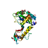
Mass: 62.068 Da / Num. of mol.: 11 / Source method: obtained synthetically / Formula: C2H6O2
#3: Water ChemComp-HOH / water / Water


Mass: 18.015 Da / Num. of mol.: 212 / Source method: isolated from a natural source / Formula: H2O

-
Experimental details

-
Experiment

ExperimentMethod: X-RAY DIFFRACTION

-
Sample preparation

CrystalDensity Matthews: 2.13 Å3/Da / Density % sol: 42 %
Crystal growDetails: 100 MM AMMONIUM ACETATE, 50 MM MAGNESIUM ACETATE, 1 MM DTT, 12-16 % PEG 6000
Crystal grow
*PLUS
Temperature: 20 ℃ / pH: 5.8 / Method: vapor diffusion, hanging drop
Components of the solutions
*PLUS
IDConc.Common nameCrystal-IDSol-ID
1100 mMammonium acetate1reservoir
250 mMmagnesium acetate1reservoir
31 mMdithiothreitol1reservoir
412-16 %PEG60001reservoir

-
Data collection

RadiationMonochromatic (M) / Laue (L): M / Scattering type: x-ray
Radiation wavelengthRelative weight: 1
ReflectionBiso Wilson estimate: 15.4 Å2
Reflection
*PLUS
Highest resolution: 1.7 Å / Lowest resolution: 20 Å / % possible obs: 86.7 % / Rmerge(I) obs: 0.03
Reflection shell
*PLUS
Highest resolution: 1.7 Å / Lowest resolution: 1.76 Å / % possible obs: 43.8 % / Rmerge(I) obs: 0.124

-
Processing

Software
NameVersionClassification
X-PLOR3.851model building
X-PLOR3.851refinement
X-PLOR3.851phasing
RefinementResolution: 1.7→8 Å / Rfactor Rfree error: 0.006 / Data cutoff high absF: 10000000 / Data cutoff low absF: 0.001 / Isotropic thermal model: RESTRAINED / Cross valid method: THROUGHOUT / σ(F): 2
RfactorNum. reflection% reflectionSelection details
Rfree0.263 2091 10 %RANDOM
Rwork0.203 ---
obs0.203 20976 85 %-
Displacement parametersBiso mean: 20 Å2
Refine analyze
FreeObs
Luzzati coordinate error0.24 Å0.19 Å
Luzzati d res low-5 Å
Luzzati sigma a0.18 Å0.19 Å
Refinement stepCycle: LAST / Resolution: 1.7→8 Å
ProteinNucleic acidLigandSolventTotal
Num. atoms1696 0 44 212 1952
Refine LS restraints
Refine-IDTypeDev idealDev ideal target
X-RAY DIFFRACTIONx_bond_d0.009
X-RAY DIFFRACTIONx_bond_d_na
X-RAY DIFFRACTIONx_bond_d_prot
X-RAY DIFFRACTIONx_angle_d
X-RAY DIFFRACTIONx_angle_d_na
X-RAY DIFFRACTIONx_angle_d_prot
X-RAY DIFFRACTIONx_angle_deg1.3
X-RAY DIFFRACTIONx_angle_deg_na
X-RAY DIFFRACTIONx_angle_deg_prot
X-RAY DIFFRACTIONx_dihedral_angle_d25
X-RAY DIFFRACTIONx_dihedral_angle_d_na
X-RAY DIFFRACTIONx_dihedral_angle_d_prot
X-RAY DIFFRACTIONx_improper_angle_d0.73
X-RAY DIFFRACTIONx_improper_angle_d_na
X-RAY DIFFRACTIONx_improper_angle_d_prot
X-RAY DIFFRACTIONx_mcbond_it21.5
X-RAY DIFFRACTIONx_mcangle_it3.082
X-RAY DIFFRACTIONx_scbond_it3.92
X-RAY DIFFRACTIONx_scangle_it6.032.5
LS refinement shellResolution: 1.7→1.81 Å / Rfactor Rfree error: 0.024 / Total num. of bins used: 6
RfactorNum. reflection% reflection
Rfree0.324 190 10.3 %
Rwork0.303 1662 -
obs--45.7 %
Xplor file
Refine-IDSerial noParam fileTopol file
X-RAY DIFFRACTION1PROTEIN_REP.PARAMTOPHCSDX.PRO
X-RAY DIFFRACTION2SEMET.PARAM
X-RAY DIFFRACTION3EDO.PARAM
Software
*PLUS
Version: 3.851 / Classification: refinement
Refinement
*PLUS
Rfactor obs: 0.208
Solvent computation
*PLUS
Displacement parameters
*PLUS
Refine LS restraints
*PLUS
Refine-IDTypeDev ideal
X-RAY DIFFRACTIONx_bond_d0.004
X-RAY DIFFRACTIONx_angle_deg1.156
X-RAY DIFFRACTIONx_dihedral_angle_d
X-RAY DIFFRACTIONx_dihedral_angle_deg25
X-RAY DIFFRACTIONx_improper_angle_d
X-RAY DIFFRACTIONx_improper_angle_deg0.73
LS refinement shell
*PLUS
Rfactor obs: 0.303

+
About Yorodumi

-
News

-
Feb 9, 2022. New format data for meta-information of EMDB entries

New format data for meta-information of EMDB entries

  • Version 3 of the EMDB header file is now the official format.
  • The previous official version 1.9 will be removed from the archive.

Related info.:EMDB header

External links:wwPDB to switch to version 3 of the EMDB data model

-
Aug 12, 2020. Covid-19 info

Covid-19 info

URL: https://pdbj.org/emnavi/covid19.php

New page: Covid-19 featured information page in EM Navigator.

Related info.:Covid-19 info / Mar 5, 2020. Novel coronavirus structure data

+
Mar 5, 2020. Novel coronavirus structure data

Novel coronavirus structure data

Related info.:Yorodumi Speices / Aug 12, 2020. Covid-19 info

External links:COVID-19 featured content - PDBj / Molecule of the Month (242):Coronavirus Proteases

+
Jan 31, 2019. EMDB accession codes are about to change! (news from PDBe EMDB page)

EMDB accession codes are about to change! (news from PDBe EMDB page)

  • The allocation of 4 digits for EMDB accession codes will soon come to an end. Whilst these codes will remain in use, new EMDB accession codes will include an additional digit and will expand incrementally as the available range of codes is exhausted. The current 4-digit format prefixed with “EMD-” (i.e. EMD-XXXX) will advance to a 5-digit format (i.e. EMD-XXXXX), and so on. It is currently estimated that the 4-digit codes will be depleted around Spring 2019, at which point the 5-digit format will come into force.
  • The EM Navigator/Yorodumi systems omit the EMD- prefix.

Related info.:Q: What is EMD? / ID/Accession-code notation in Yorodumi/EM Navigator

External links:EMDB Accession Codes are Changing Soon! / Contact to PDBj

+
Jul 12, 2017. Major update of PDB

Major update of PDB

  • wwPDB released updated PDB data conforming to the new PDBx/mmCIF dictionary.
  • This is a major update changing the version number from 4 to 5, and with Remediation, in which all the entries are updated.
  • In this update, many items about electron microscopy experimental information are reorganized (e.g. em_software).
  • Now, EM Navigator and Yorodumi are based on the updated data.

External links:wwPDB Remediation / Enriched Model Files Conforming to OneDep Data Standards Now Available in the PDB FTP Archive

-
Yorodumi

Thousand views of thousand structures

  • Yorodumi is a browser for structure data from EMDB, PDB, SASBDB, etc.
  • This page is also the successor to EM Navigator detail page, and also detail information page/front-end page for Omokage search.
  • The word "yorodu" (or yorozu) is an old Japanese word meaning "ten thousand". "mi" (miru) is to see.

Related info.:EMDB / PDB / SASBDB / Comparison of 3 databanks / Yorodumi Search / Aug 31, 2016. New EM Navigator & Yorodumi / Yorodumi Papers / Jmol/JSmol / Function and homology information / Changes in new EM Navigator and Yorodumi

Read more