 Movie
Movie Controller


+ Open data

- Basic information
| Entry | Database: PDB / ID: 2cuo | ||||||
|---|---|---|---|---|---|---|---|
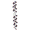
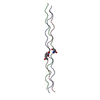
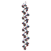
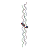
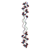
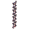
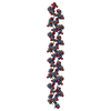
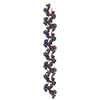
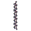
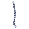
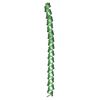
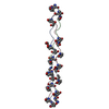
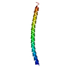
| Title | Collagen model peptide (PRO-PRO-GLY)9 | ||||||
 Components Components | COLLAGEN MODEL PEPTIDE (PRO-PRO-GLY)9 | ||||||
 Keywords Keywords | STRUCTURAL PROTEIN / COLLAGEN MODEL PEPTIDE / TRIPLE-HELIX / PUCKERING | ||||||
| Function / homology | Saimiri transformation-associated protein / Collagen triple helix repeat / Collagen triple helix repeat (20 copies) / membrane / Saimiri transformation-associated protein Function and homology information Function and homology information | ||||||
| Method |  X-RAY DIFFRACTION / SYNCHROTRON / MOLECULAR REPLACEMENT / Resolution: 1.33 Å X-RAY DIFFRACTION / SYNCHROTRON / MOLECULAR REPLACEMENT / Resolution: 1.33 Å | ||||||
 Authors Authors | Hongo, C. / Noguchi, K. / Okuyama, K. / Tanaka, Y. / Nishino, N. | ||||||
 Citation Citation | Journal: J.Biochem.(Tokyo) / Year: 2005 Title: Repetitive interactions observed in the crystal structure of a collagen-model peptide, [(Pro-Pro-Gly)9]3 Authors: Hongo, C. / Noguchi, K. / Okuyama, K. / Tanaka, Y. / Nishino, N. | ||||||
| History |
|
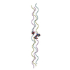
- Structure visualization
| Structure viewer | Molecule: MolmilJmol/JSmol |
|---|
- Downloads & links
-Download
| PDBx/mmCIF format | 2cuo.cif.gz | 61 KB | Display | PDBx/mmCIF format |
|---|---|---|---|---|
| PDB format | pdb2cuo.ent.gz | 48.3 KB | Display | PDB format |
| PDBx/mmJSON format | 2cuo.json.gz | Tree view | PDBx/mmJSON format | |
| Others |  Other downloads Other downloads |
-Validation report
| Summary document | 2cuo_validation.pdf.gz | 371.4 KB | Display | wwPDB validaton report |
|---|---|---|---|---|
| Full document | 2cuo_full_validation.pdf.gz | 371.8 KB | Display | |
| Data in XML | 2cuo_validation.xml.gz | 3.6 KB | Display | |
| Data in CIF | 2cuo_validation.cif.gz | 7.2 KB | Display | |
| Arichive directory | https://data.pdbj.org/pub/pdb/validation_reports/cu/2cuoftp://data.pdbj.org/pub/pdb/validation_reports/cu/2cuo | HTTPS FTP |
-Related structure data
| Related structure data | 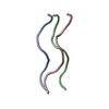 1ittS S: Starting model for refinement |
|---|---|
| Similar structure data |
-Links
 PDBj
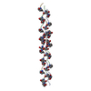
PDBj- Assembly
| Deposited unit | 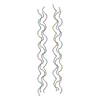
| ||||||||
|---|---|---|---|---|---|---|---|---|---|
| 1 | 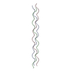
| ||||||||
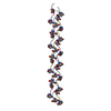
| 2 | 
| ||||||||
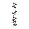
| 3 |
| ||||||||
| Unit cell |
|
-Components
| #1: Protein/peptide | Mass: 2279.547 Da / Num. of mol.: 6 / Source method: obtained synthetically / Details: COLLAGEN MODEL PEPTIDE WAS CHEMICALLY SYSTHESIZED / References: UniProt: Q80BK4*PLUS #2: Water | ChemComp-HOH / |  Mass: 18.015 Da / Num. of mol.: 332 / Source method: isolated from a natural source / Formula: H2O Mass: 18.015 Da / Num. of mol.: 332 / Source method: isolated from a natural source / Formula: H2O |
|---|
-Experimental details
-Experiment
| Experiment | Method: X-RAY DIFFRACTION / Number of used crystals: 1 |
|---|
- Sample preparation
| Crystal | Density Matthews: 1.71 Å3/Da / Density % sol: 27.9 % |
|---|---|
| Crystal grow | Temperature: 277 K / Method: vapor diffusion, hanging drop Details: PEG400, ACETIC ACID, SODIUM AZIDE, VAPOR DIFFUSION, HANGING DROP, temperature 277K |
-Data collection
| Diffraction | Mean temperature: 100 K |
|---|---|
| Diffraction source | Source: SYNCHROTRON / Site: SPring-8  / Beamline: BL40B2 / Wavelength: 1 / Wavelength: 1 Å / Beamline: BL40B2 / Wavelength: 1 / Wavelength: 1 Å |
| Detector | Type: ADSC QUANTUM 4r / Detector: CCD / Date: Jun 20, 2001 / Details: 1-M-LONG BENT-CYLINDER MIRROR |
| Radiation | Monochromator: FIXED-EXIT DOUBLE CRYSTAL / Protocol: SINGLE WAVELENGTH / Monochromatic (M) / Laue (L): M / Scattering type: x-ray |
| Radiation wavelength | Wavelength: 1 Å / Relative weight: 1 |
| Reflection | Resolution: 1.33→25.21 Å / Num. obs: 24336 / % possible obs: 97.7 % / Observed criterion σ(F): 1 / Observed criterion σ(I): 1 / Redundancy: 3.2 % / Rmerge(I) obs: 0.07 / Net I/σ(I): 4.8 |
| Reflection shell | Resolution: 1.33→1.38 Å / Redundancy: 3.3 % / Rmerge(I) obs: 0.269 / Mean I/σ(I) obs: 1.8 / % possible all: 98.3 |
- Processing
| Software |
| |||||||||||||||||||||||||||||||||
|---|---|---|---|---|---|---|---|---|---|---|---|---|---|---|---|---|---|---|---|---|---|---|---|---|---|---|---|---|---|---|---|---|---|---|
| Refinement | Method to determine structure: MOLECULAR REPLACEMENT Starting model: PDB ENTRY 1ITT Resolution: 1.33→8 Å / Num. parameters: 9953 / Num. restraintsaints: 12321 / σ(F): 1 / Stereochemistry target values: ENGH AND HUBER / Details: ANISOTROPIC REFINEMENT REDUCED FREE R
| |||||||||||||||||||||||||||||||||
| Solvent computation | Solvent model: MOEWS & KRETSINGER, J.MOL.BIOL.91(1973)201-228 | |||||||||||||||||||||||||||||||||
| Refine analyze | Num. disordered residues: 0 / Occupancy sum hydrogen: 918 / Occupancy sum non hydrogen: 1290 | |||||||||||||||||||||||||||||||||
| Refinement step | Cycle: LAST / Resolution: 1.33→8 Å
| |||||||||||||||||||||||||||||||||
| Refine LS restraints |
|
