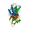
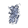
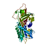
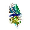
SHEET THE SHEET STRUCTURE OF THIS MOLECULE IS BIFURCATED. IN ORDER TO REPRESENT THIS FEATURE IN ... SHEET THE SHEET STRUCTURE OF THIS MOLECULE IS BIFURCATED. IN ORDER TO REPRESENT THIS FEATURE IN THE SHEET RECORDS BELOW, TWO SHEETS ARE DEFINED.
FIRST 18 RESIDUES OF MATURE SEQUENCE REPLACED BY HIS-TAG AND THROMBIN CLEAVAGE SITE. GLY-SER LEFT ...FIRST 18 RESIDUES OF MATURE SEQUENCE REPLACED BY HIS-TAG AND THROMBIN CLEAVAGE SITE. GLY-SER LEFT AT N-TERMINUS AFTER CLEAVAGE.
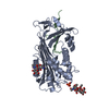
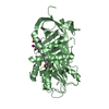
解像度: 2.8→30.14 Å / Cor.coef. Fo:Fc: 0.918 / Cor.coef. Fo:Fc free: 0.879 / SU B: 41.661 / SU ML: 0.375 / TLS residual ADP flag: LIKELY RESIDUAL / 交差検証法: THROUGHOUT / ESU R Free: 0.469 / 立体化学のターゲット値: MAXIMUM LIKELIHOOD 詳細: HYDROGENS HAVE BEEN ADDED IN THE RIDING POSITIONS. RESIDUES 17-19, 350-357 AND 395 OF BOTH CHAINS A AND B ARE DISORDERED
Rfactor
反射数
%反射
Selection details
Rfree
0.283
972
5.1 %
RANDOM
Rwork
0.235
-
-
-
obs
0.237
18166
93.6 %
-
溶媒の処理
イオンプローブ半径: 0.8 Å / 減衰半径: 0.8 Å / VDWプローブ半径: 1.4 Å / 溶媒モデル: MASK
 ムービー
ムービー コントローラー


 データを開く
 基本情報
基本情報 要素
要素 キーワード
キーワード 機能・相同性情報
 HOMO SAPIENS (ヒト)
 X線回折 /
  データ登録者
 引用
引用 構造の表示
構造の表示 ダウンロードとリンク
 その他のダウンロード


 PDBj
PDBj
 集合体
集合体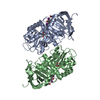

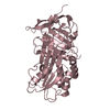
 分子量: 776.870 Da / 分子数: 2 / 由来タイプ: 合成 / 式: C15H11I4NO4 / コメント: ホルモン*YM
 分子量: 92.094 Da / 分子数: 2 / 由来タイプ: 合成 / 式: C3H8O3
 分子量: 18.015 Da / 分子数: 26 / 由来タイプ: 天然 / 式: H2O
 試料調製
試料調製 / ビームライン: PX14.2 / 波長: 0.979
  解析
解析