 Movie
Movie Controller


+ Open data

- Basic information
| Entry | Database: PDB / ID: 1psi | ||||||
|---|---|---|---|---|---|---|---|
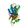
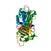
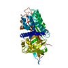
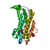
| Title | Intact recombined alpha1-antitrypsin mutant PHE 51 to LEU | ||||||
 Components Components | ALPHA=1=-ANTITRYPSIN | ||||||
 Keywords Keywords | SERINE PROTEASE INHIBITOR / SERPIN / GLYCOPROTEIN / POLYMORPHISM / EMPHYSEMA / DISEASE MUTATION / ACUTE PHASE | ||||||
| Function / homology |  Function and homology information Function and homology informationCargo concentration in the ER / COPII-coated ER to Golgi transport vesicle / COPII-mediated vesicle transport / endoplasmic reticulum-Golgi intermediate compartment membrane / platelet alpha granule lumen / acute-phase response / Post-translational protein phosphorylation / serine-type endopeptidase inhibitor activity / Regulation of Insulin-like Growth Factor (IGF) transport and uptake by Insulin-like Growth Factor Binding Proteins (IGFBPs) / blood coagulation ...Cargo concentration in the ER / COPII-coated ER to Golgi transport vesicle / COPII-mediated vesicle transport / endoplasmic reticulum-Golgi intermediate compartment membrane / platelet alpha granule lumen / acute-phase response / Post-translational protein phosphorylation / serine-type endopeptidase inhibitor activity / Regulation of Insulin-like Growth Factor (IGF) transport and uptake by Insulin-like Growth Factor Binding Proteins (IGFBPs) / blood coagulation / Platelet degranulation / extracellular matrix / protease binding / ficolin-1-rich granule lumen / endoplasmic reticulum lumen / Neutrophil degranulation / Golgi apparatus / endoplasmic reticulum / : / extracellular exosome / extracellular region / identical protein binding Similarity search - Function | ||||||
| Biological species |  Homo sapiens (human) Homo sapiens (human) | ||||||
| Method |  X-RAY DIFFRACTION / MOLECULAR REPLACEMENT / Resolution: 2.92 Å X-RAY DIFFRACTION / MOLECULAR REPLACEMENT / Resolution: 2.92 Å | ||||||
 Authors Authors | Abrahams, J.P. / Elliott, P.R. / Lomas, D.A. / Carrell, R.W. | ||||||
 Citation Citation | Journal: Nat.Struct.Biol. / Year: 1996 Title: Inhibitory conformation of the reactive loop of alpha 1-antitrypsin. Authors: Elliott, P.R. / Lomas, D.A. / Carrell, R.W. / Abrahams, J.P. | ||||||
| History |
|
- Structure visualization
| Structure viewer | Molecule: MolmilJmol/JSmol |
|---|
- Downloads & links
-Download
| PDBx/mmCIF format | 1psi.cif.gz | 81 KB | Display | PDBx/mmCIF format |
|---|---|---|---|---|
| PDB format | pdb1psi.ent.gz | 63.8 KB | Display | PDB format |
| PDBx/mmJSON format | 1psi.json.gz | Tree view | PDBx/mmJSON format | |
| Others |  Other downloads Other downloads |
-Validation report
| Arichive directory | https://data.pdbj.org/pub/pdb/validation_reports/ps/1psiftp://data.pdbj.org/pub/pdb/validation_reports/ps/1psi | HTTPS FTP |
|---|
-Related structure data
| Related structure data | 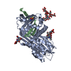 9apiS S: Starting model for refinement |
|---|---|
| Similar structure data |


-Links
 PDBj
PDBj

- Assembly
| Deposited unit | 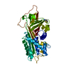
| ||||||||
|---|---|---|---|---|---|---|---|---|---|
| 1 |
| ||||||||
| Unit cell |
|
-Components
| #1: Protein | Mass: 44406.539 Da / Num. of mol.: 1 / Mutation: F51L Source method: isolated from a genetically manipulated source Source: (gene. exp.) Homo sapiens (human) / Tissue: BLOOD PLASMA / Gene: ALPHA-1-ANTITRYPSIN / Organ: BLOOD / Plasmid: PTERMAT / Gene (production host): ALPHA-1-ANTITRYPSIN / Production host:  |
|---|
-Experimental details
-Experiment
| Experiment | Method: X-RAY DIFFRACTION / Number of used crystals: 1 |
|---|
- Sample preparation
| Crystal | Density Matthews: 2.11 Å3/Da / Density % sol: 45 % | |||||||||||||||||||||||||||||||||||
|---|---|---|---|---|---|---|---|---|---|---|---|---|---|---|---|---|---|---|---|---|---|---|---|---|---|---|---|---|---|---|---|---|---|---|---|---|
| Crystal grow | pH: 6 Details: 24% PEG, 0.2 M SODIUM ACETATE, 0.1 M TRIS.HCL, PH 6.0, 0.002 M FESO4, 25 DEG CELSIUS | |||||||||||||||||||||||||||||||||||
| Crystal grow | *PLUS Temperature: 18 ℃ / Method: vapor diffusion, hanging drop | |||||||||||||||||||||||||||||||||||
| Components of the solutions | *PLUS
|
-Data collection
| Diffraction | Mean temperature: 100 K |
|---|---|
| Diffraction source | Source: ROTATING ANODE / Type: ELLIOTT GX-11 / Wavelength: 1.5418 |
| Detector | Type: MARRESEARCH / Detector: IMAGE PLATE / Date: Feb 12, 1996 / Details: NICKEL COATED DOUBLE MIRROR |
| Radiation | Monochromatic (M) / Laue (L): M / Scattering type: x-ray |
| Radiation wavelength | Wavelength: 1.5418 Å / Relative weight: 1 |
| Reflection | Resolution: 2.92→19.5 Å / Num. obs: 7651 / % possible obs: 91.7 % / Observed criterion σ(I): 3.5 / Redundancy: 2.2 % / Rmerge(I) obs: 0.114 / Rsym value: 0.114 / Net I/σ(I): 8.36 |
| Reflection shell | Resolution: 2.91→3.11 Å / Redundancy: 2 % / Rmerge(I) obs: 0.204 / Mean I/σ(I) obs: 4 / Rsym value: 0.204 / % possible all: 74.4 |
| Reflection | *PLUS Highest resolution: 2.9 Å / Lowest resolution: 6 Å / Rmerge(I) obs: 0.114 |
| Reflection shell | *PLUS Highest resolution: 2.9 Å / Lowest resolution: 3.1 Å / % possible obs: 74.4 % / Rmerge(I) obs: 0.204 |
- Processing
| Software |
| ||||||||||||||||||||||||||||||||||||||||||||||||||||||||||||
|---|---|---|---|---|---|---|---|---|---|---|---|---|---|---|---|---|---|---|---|---|---|---|---|---|---|---|---|---|---|---|---|---|---|---|---|---|---|---|---|---|---|---|---|---|---|---|---|---|---|---|---|---|---|---|---|---|---|---|---|---|---|
| Refinement | Method to determine structure: MOLECULAR REPLACEMENT Starting model: CLEAVED ALPHA1-ANTITRYPSIN (PDB ENTRY 9API) Resolution: 2.92→6 Å / σ(F): 0 Details: RESIDUES 106 - 108 ARE POORLY ORDERED. ALTHOUGH SOME CONNECTED DENSITY EXISTS, A STEREOCHEMICALLY CORRECT MODEL FITTING THE DATA COULD NOT BE BUILT. THE OCCUPANCIES OF THESE RESIDUES ARE SET ...Details: RESIDUES 106 - 108 ARE POORLY ORDERED. ALTHOUGH SOME CONNECTED DENSITY EXISTS, A STEREOCHEMICALLY CORRECT MODEL FITTING THE DATA COULD NOT BE BUILT. THE OCCUPANCIES OF THESE RESIDUES ARE SET TO ZERO. PRO 106 - SER 108 IS A POORLY ORDERED LOOP, PRESENT IN MULTIPLE CONFORMATIONS. A STEREOCHEMICALLY VALID MODEL WAS BUILT, BUT THE OCCUPANCIES ARE SET TO ZERO.
| ||||||||||||||||||||||||||||||||||||||||||||||||||||||||||||
| Refinement step | Cycle: LAST / Resolution: 2.92→6 Å
| ||||||||||||||||||||||||||||||||||||||||||||||||||||||||||||
| Refine LS restraints |
| ||||||||||||||||||||||||||||||||||||||||||||||||||||||||||||
| Software | *PLUS Name: 'TNT and X-PLOR' / Classification: refinement | ||||||||||||||||||||||||||||||||||||||||||||||||||||||||||||
| Refinement | *PLUS % reflection Rfree: 10 % / Rfactor obs: 0.218 / Rfactor Rfree: 0.288 / Rfactor Rwork: 0.215 / Highest resolution: 2.9 Å / Num. reflection all: 6699 / Num. reflection Rfree: 754 | ||||||||||||||||||||||||||||||||||||||||||||||||||||||||||||
| Solvent computation | *PLUS | ||||||||||||||||||||||||||||||||||||||||||||||||||||||||||||
| Displacement parameters | *PLUS | ||||||||||||||||||||||||||||||||||||||||||||||||||||||||||||
| Refine LS restraints | *PLUS
|
