 Movie
Movie Controller

[English] 日本語
 Yorodumi
- PDB-2byp: Crystal structure of Aplysia californica AChBP in complex with al... -
+ Open data

- Basic information
| Entry | Database: PDB / ID: 2byp | ||||||
|---|---|---|---|---|---|---|---|
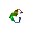
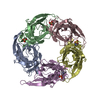
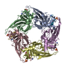
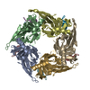
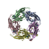
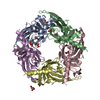
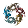
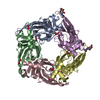
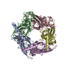
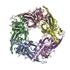
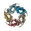
| Title | Crystal structure of Aplysia californica AChBP in complex with alpha- conotoxin ImI | ||||||
 Components Components |
| ||||||
 Keywords Keywords | RECEPTOR / RECEPTOR COMPLEX / NICOTINIC ACETYLCHOLINE RECEPTOR COMPLEX / CONOTOXIN | ||||||
| Function / homology |  Function and homology information Function and homology informationhost cell postsynaptic membrane / acetylcholine receptor inhibitor activity / ion channel regulator activity / extracellular ligand-gated monoatomic ion channel activity / transmembrane signaling receptor activity / toxin activity / extracellular region / membrane / metal ion binding / identical protein binding Similarity search - Function | ||||||
| Biological species |  CONUS IMPERIALIS (invertebrata) CONUS IMPERIALIS (invertebrata) | ||||||
| Method |  X-RAY DIFFRACTION / SYNCHROTRON / MOLECULAR REPLACEMENT / Resolution: 2.07 Å X-RAY DIFFRACTION / SYNCHROTRON / MOLECULAR REPLACEMENT / Resolution: 2.07 Å | ||||||
 Authors Authors | Hansen, S.B. / Sulzenbacher, G. / Huxford, T. / Marchot, P. / Taylor, P. / Bourne, Y. | ||||||
 Citation Citation | Journal: Embo J. / Year: 2005 Title: Structures of Aplysia Achbp Complexes with Nicotinic Agonists and Antagonists Reveal Distinctive Binding Interfaces and Conformations. Authors: Hansen, S.B. / Sulzenbacher, G. / Huxford, T. / Marchot, P. / Taylor, P. / Bourne, Y. | ||||||
| History |
| ||||||
| Remark 700 | SHEET THE SHEET STRUCTURE OF THIS MOLECULE IS BIFURCATED. IN ORDER TO REPRESENT THIS FEATURE IN ... SHEET THE SHEET STRUCTURE OF THIS MOLECULE IS BIFURCATED. IN ORDER TO REPRESENT THIS FEATURE IN THE SHEET RECORDS BELOW, TWO SHEETS ARE DEFINED. |
- Structure visualization
| Structure viewer | Molecule: MolmilJmol/JSmol |
|---|
- Downloads & links
-Download
| PDBx/mmCIF format | 2byp.cif.gz | 256.1 KB | Display | PDBx/mmCIF format |
|---|---|---|---|---|
| PDB format | pdb2byp.ent.gz | 210.3 KB | Display | PDB format |
| PDBx/mmJSON format | 2byp.json.gz | Tree view | PDBx/mmJSON format | |
| Others |  Other downloads Other downloads |
-Validation report
| Arichive directory | https://data.pdbj.org/pub/pdb/validation_reports/by/2bypftp://data.pdbj.org/pub/pdb/validation_reports/by/2byp | HTTPS FTP |
|---|
-Related structure data
| Related structure data | 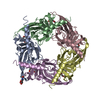 2bynSC 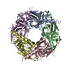 2byqC 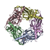 2byrC 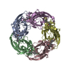 2bysC S: Starting model for refinement C: citing same article ( |
|---|---|
| Similar structure data |


-Links
 PDBj
PDBj

- Assembly
| Deposited unit | 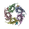
| ||||||||||||||||||
|---|---|---|---|---|---|---|---|---|---|---|---|---|---|---|---|---|---|---|---|
| 1 |
| ||||||||||||||||||
| Unit cell |
| ||||||||||||||||||
| Components on special symmetry positions |
|
-Components
| #1: Protein | Mass: 24320.039 Da / Num. of mol.: 5 Source method: isolated from a genetically manipulated source Source: (gene. exp.) Cell: SENSORY CELL / Cell line (production host): HEK293 / Production host:  HOMO SAPIENS (human) / Tissue (production host): KIDNEY / References: UniProt: Q8WSF8 HOMO SAPIENS (human) / Tissue (production host): KIDNEY / References: UniProt: Q8WSF8#2: Protein/peptide | Mass: 1357.609 Da / Num. of mol.: 5 / Source method: obtained synthetically / Source: (synth.) CONUS IMPERIALIS (invertebrata) / References: UniProt: P50983#3: Sugar | ChemComp-NAG / |   Type: D-saccharide, beta linking / Mass: 221.208 Da / Num. of mol.: 1 Type: D-saccharide, beta linking / Mass: 221.208 Da / Num. of mol.: 1Source method: isolated from a genetically manipulated source Formula: C8H15NO6 #4: Water | ChemComp-HOH / |  Mass: 18.015 Da / Num. of mol.: 1042 / Source method: isolated from a natural source / Formula: H2O Mass: 18.015 Da / Num. of mol.: 1042 / Source method: isolated from a natural source / Formula: H2O |
|---|
-Experimental details
-Experiment
| Experiment | Method: X-RAY DIFFRACTION / Number of used crystals: 1 |
|---|
- Sample preparation
| Crystal | Density Matthews: 2.44 Å3/Da / Density % sol: 49.25 % |
|---|---|
| Crystal grow | pH: 7.5 / Details: 11-14% PEG-4000, 0.1 M TRIS, PH 7.5, 0.4 M MGCL2 |
-Data collection
| Diffraction | Mean temperature: 100 K |
|---|---|
| Diffraction source | Source: SYNCHROTRON / Site: ALS  / Beamline: 8.2.1 / Wavelength: 0.976 / Beamline: 8.2.1 / Wavelength: 0.976 |
| Radiation | Protocol: SINGLE WAVELENGTH / Monochromatic (M) / Laue (L): M / Scattering type: x-ray |
| Radiation wavelength | Wavelength: 0.976 Å / Relative weight: 1 |
| Reflection | Resolution: 2.07→50 Å / Num. obs: 86601 / % possible obs: 99.9 % / Observed criterion σ(I): 0 / Redundancy: 5.1 % / Rmerge(I) obs: 0.08 / Net I/σ(I): 29.5 |
- Processing
| Software |
| ||||||||||||||||||||||||||||||||||||||||||||||||||||||||||||||||||||||||||||||||||||||||||||||||||||||||||||||||||||||||||||||||||||||||||||||||||||||||||||||||||||||||||||||||||||||
|---|---|---|---|---|---|---|---|---|---|---|---|---|---|---|---|---|---|---|---|---|---|---|---|---|---|---|---|---|---|---|---|---|---|---|---|---|---|---|---|---|---|---|---|---|---|---|---|---|---|---|---|---|---|---|---|---|---|---|---|---|---|---|---|---|---|---|---|---|---|---|---|---|---|---|---|---|---|---|---|---|---|---|---|---|---|---|---|---|---|---|---|---|---|---|---|---|---|---|---|---|---|---|---|---|---|---|---|---|---|---|---|---|---|---|---|---|---|---|---|---|---|---|---|---|---|---|---|---|---|---|---|---|---|---|---|---|---|---|---|---|---|---|---|---|---|---|---|---|---|---|---|---|---|---|---|---|---|---|---|---|---|---|---|---|---|---|---|---|---|---|---|---|---|---|---|---|---|---|---|---|---|---|---|
| Refinement | Method to determine structure: MOLECULAR REPLACEMENT Starting model: PDB ENTRY 2BYN Resolution: 2.07→20 Å / Cor.coef. Fo:Fc: 0.964 / Cor.coef. Fo:Fc free: 0.949 / SU B: 7.782 / SU ML: 0.122 / TLS residual ADP flag: UNVERIFIED / Cross valid method: THROUGHOUT / ESU R: 0.176 / ESU R Free: 0.155 / Stereochemistry target values: MAXIMUM LIKELIHOOD / Details: HYDROGENS HAVE BEEN ADDED IN THE RIDING POSITIONS.
| ||||||||||||||||||||||||||||||||||||||||||||||||||||||||||||||||||||||||||||||||||||||||||||||||||||||||||||||||||||||||||||||||||||||||||||||||||||||||||||||||||||||||||||||||||||||
| Solvent computation | Ion probe radii: 0.8 Å / Shrinkage radii: 0.8 Å / VDW probe radii: 1.2 Å / Solvent model: MASK | ||||||||||||||||||||||||||||||||||||||||||||||||||||||||||||||||||||||||||||||||||||||||||||||||||||||||||||||||||||||||||||||||||||||||||||||||||||||||||||||||||||||||||||||||||||||
| Displacement parameters | Biso mean: 38.98 Å2
| ||||||||||||||||||||||||||||||||||||||||||||||||||||||||||||||||||||||||||||||||||||||||||||||||||||||||||||||||||||||||||||||||||||||||||||||||||||||||||||||||||||||||||||||||||||||
| Refinement step | Cycle: LAST / Resolution: 2.07→20 Å
| ||||||||||||||||||||||||||||||||||||||||||||||||||||||||||||||||||||||||||||||||||||||||||||||||||||||||||||||||||||||||||||||||||||||||||||||||||||||||||||||||||||||||||||||||||||||
| Refine LS restraints |
|