 Movie
Movie Controller

[English] 日本語
 Yorodumi
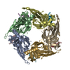
- PDB-2br8: Crystal Structure of Acetylcholine-binding Protein (AChBP) from A... -
+ Open data

- Basic information
| Entry | Database: PDB / ID: 2br8 | ||||||
|---|---|---|---|---|---|---|---|
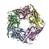
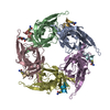
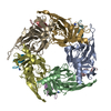
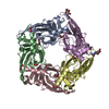
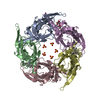
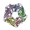
| Title | Crystal Structure of Acetylcholine-binding Protein (AChBP) from Aplysia californica in complex with an alpha-conotoxin PnIA variant | ||||||
 Components Components |
| ||||||
 Keywords Keywords | RECEPTOR/INHIBITOR / RECEPTOR-INHIBITOR COMPLEX / GLYCOPROTEIN / IGG-FOLD / IMMUNOGLOBULIN DOMAIN / PENTAMER / NICOTINIC RECEPTOR / ALPHA-CONOTOXIN / RECEPTOR / ACETYLCHOLINE RECEPTOR INHIBITOR / AMIDATION / NEUROTOXIN / POSTSYNAPTIC NEUROTOXIN / SULFATION / TOXIN | ||||||
| Function / homology |  Function and homology information Function and homology informationhost cell postsynaptic membrane / acetylcholine receptor inhibitor activity / ion channel regulator activity / extracellular ligand-gated monoatomic ion channel activity / transmembrane signaling receptor activity / toxin activity / extracellular region / membrane / metal ion binding / identical protein binding Similarity search - Function | ||||||
| Biological species |  CONUS PENNACEUS (invertebrata) CONUS PENNACEUS (invertebrata) | ||||||
| Method |  X-RAY DIFFRACTION / SYNCHROTRON / MOLECULAR REPLACEMENT / Resolution: 2.4 Å X-RAY DIFFRACTION / SYNCHROTRON / MOLECULAR REPLACEMENT / Resolution: 2.4 Å | ||||||
 Authors Authors | Celie, P.H.N. / Kasheverov, I.E. / Mordvintsev, D.Y. / Hogg, R.C. / van Nierop, P. / van Elk, R. / van Rossum-Fikkert, S.E. / Zhmak, M.N. / Bertrand, D. / Tsetlin, V. ...Celie, P.H.N. / Kasheverov, I.E. / Mordvintsev, D.Y. / Hogg, R.C. / van Nierop, P. / van Elk, R. / van Rossum-Fikkert, S.E. / Zhmak, M.N. / Bertrand, D. / Tsetlin, V. / Sixma, T.K. / Smit, A.B. | ||||||
 Citation Citation | Journal: Nat.Struct.Mol.Biol. / Year: 2005 Title: Crystal Structure of Nicotinic Acetylcholine Receptor Homolog Achbp in Complex with an Alpha-Conotoxin Pnia Variant Authors: Celie, P.H.N. / Kasheverov, I.E. / Mordvintsev, D.Y. / Hogg, R.C. / Van Nierop, P. / Van Elk, R. / Van Rossum-Fikkert, S.E. / Zhmak, M.N. / Bertrand, D. / Tsetlin, V. / Sixma, T.K. / Smit, A.B. | ||||||
| History |
| ||||||
| Remark 700 | SHEET THE SHEET STRUCTURE OF THIS MOLECULE IS BIFURCATED. IN ORDER TO REPRESENT THIS FEATURE IN ... SHEET THE SHEET STRUCTURE OF THIS MOLECULE IS BIFURCATED. IN ORDER TO REPRESENT THIS FEATURE IN THE SHEET RECORDS BELOW, TWO SHEETS ARE DEFINED. |
- Structure visualization
| Structure viewer | Molecule: MolmilJmol/JSmol |
|---|
- Downloads & links
-Download
| PDBx/mmCIF format | 2br8.cif.gz | 225 KB | Display | PDBx/mmCIF format |
|---|---|---|---|---|
| PDB format | pdb2br8.ent.gz | 184.1 KB | Display | PDB format |
| PDBx/mmJSON format | 2br8.json.gz | Tree view | PDBx/mmJSON format | |
| Others |  Other downloads Other downloads |
-Validation report
| Arichive directory | https://data.pdbj.org/pub/pdb/validation_reports/br/2br8ftp://data.pdbj.org/pub/pdb/validation_reports/br/2br8 | HTTPS FTP |
|---|
-Related structure data
| Related structure data | 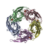 2br7C 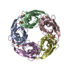 1ux2S C: citing same article ( S: Starting model for refinement |
|---|---|
| Similar structure data |


-Links
 PDBj
PDBj

- Assembly
| Deposited unit | 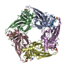
| ||||||||||||||||||||||||||||||||||||
|---|---|---|---|---|---|---|---|---|---|---|---|---|---|---|---|---|---|---|---|---|---|---|---|---|---|---|---|---|---|---|---|---|---|---|---|---|---|
| 1 |
| ||||||||||||||||||||||||||||||||||||
| Unit cell |
| ||||||||||||||||||||||||||||||||||||
| Noncrystallographic symmetry (NCS) | NCS oper:
|
-Components
| #1: Protein | Mass: 24688.578 Da / Num. of mol.: 5 Source method: isolated from a genetically manipulated source Details: HEPES BUFFER MOLECULE IDENTIFIED IN 4 OUT OF FIVE BINDING SITES Source: (gene. exp.) Cell: GLIAL CELL / Plasmid: PFASTBACI / Cell line (production host): SF9 / Production host:   SPODOPTERA FRUGIPERDA (fall armyworm) / References: UniProt: Q8WSF8 SPODOPTERA FRUGIPERDA (fall armyworm) / References: UniProt: Q8WSF8#2: Protein/peptide | Mass: 1682.021 Da / Num. of mol.: 5 / Mutation: YES / Source method: obtained synthetically / Source: (synth.) CONUS PENNACEUS (invertebrata) / References: UniProt: P50984#3: Chemical | ChemComp-SO4 /   Mass: 96.063 Da / Num. of mol.: 5 / Source method: obtained synthetically / Formula: SO4 Mass: 96.063 Da / Num. of mol.: 5 / Source method: obtained synthetically / Formula: SO4#4: Water | ChemComp-HOH / |  Mass: 18.015 Da / Num. of mol.: 235 / Source method: isolated from a natural source / Formula: H2O Mass: 18.015 Da / Num. of mol.: 235 / Source method: isolated from a natural source / Formula: H2OCompound details | FUNCTION: ALPHA-CONOTOXINS INHIBIT THE NICOTINIC ACETYLCHOLINE RECEPTORS (NACHR) ENGINEERED RESIDUE ...FUNCTION: ALPHA-CONOTOXINS | Has protein modification | Y | Sequence details | PROTEIN CONTAINS SIGNAL SEQUENCE MLVSVYLALLVACVGQAHS THAT IS CLEAVED UPON SECRETION AND IS NOT ...PROTEIN CONTAINS SIGNAL SEQUENCE MLVSVYLALL | |
|---|
-Experimental details
-Experiment
| Experiment | Method: X-RAY DIFFRACTION / Number of used crystals: 1 |
|---|
- Sample preparation
| Crystal | Density Matthews: 2.9 Å3/Da / Density % sol: 57.5 % |
|---|---|
| Crystal grow | pH: 7.5 Details: 18 % PEG 3350, 180 MM NA2SO4, 100 MM BIS-TRIS PROPANE PH 7.5 |
-Data collection
| Diffraction | Mean temperature: 100 K |
|---|---|
| Diffraction source | Source: SYNCHROTRON / Site: ESRF  / Beamline: ID14-2 / Wavelength: 0.933 / Beamline: ID14-2 / Wavelength: 0.933 |
| Detector | Type: ADSC CCD / Detector: CCD / Date: Sep 30, 2004 |
| Radiation | Protocol: SINGLE WAVELENGTH / Monochromatic (M) / Laue (L): M / Scattering type: x-ray |
| Radiation wavelength | Wavelength: 0.933 Å / Relative weight: 1 |
| Reflection | Resolution: 2.4→50 Å / Num. obs: 55263 / % possible obs: 97.9 % / Observed criterion σ(I): 1.8 / Redundancy: 3.45 % / Rmerge(I) obs: 0.1 / Net I/σ(I): 6 |
| Reflection shell | Resolution: 2.4→2.52 Å / Redundancy: 2.98 % / Rmerge(I) obs: 0.4 / Mean I/σ(I) obs: 1.8 / % possible all: 90.4 |
- Processing
| Software |
| ||||||||||||||||||||||||||||||||||||||||||||||||||||||||||||||||||||||||||||||||||||||||||||||||||||||||||||||||||||||||||||||||||||||||||||||||||||||||||||||||||||||||||||||||||||||
|---|---|---|---|---|---|---|---|---|---|---|---|---|---|---|---|---|---|---|---|---|---|---|---|---|---|---|---|---|---|---|---|---|---|---|---|---|---|---|---|---|---|---|---|---|---|---|---|---|---|---|---|---|---|---|---|---|---|---|---|---|---|---|---|---|---|---|---|---|---|---|---|---|---|---|---|---|---|---|---|---|---|---|---|---|---|---|---|---|---|---|---|---|---|---|---|---|---|---|---|---|---|---|---|---|---|---|---|---|---|---|---|---|---|---|---|---|---|---|---|---|---|---|---|---|---|---|---|---|---|---|---|---|---|---|---|---|---|---|---|---|---|---|---|---|---|---|---|---|---|---|---|---|---|---|---|---|---|---|---|---|---|---|---|---|---|---|---|---|---|---|---|---|---|---|---|---|---|---|---|---|---|---|---|
| Refinement | Method to determine structure: MOLECULAR REPLACEMENT Starting model: PDB ENTRY 1UX2 Resolution: 2.4→19.8 Å / Cor.coef. Fo:Fc: 0.942 / Cor.coef. Fo:Fc free: 0.907 / SU B: 18.569 / SU ML: 0.209 / Cross valid method: THROUGHOUT / ESU R: 0.368 / ESU R Free: 0.256 / Stereochemistry target values: MAXIMUM LIKELIHOOD / Details: HYDROGENS HAVE BEEN ADDED IN THE RIDING POSITIONS.
| ||||||||||||||||||||||||||||||||||||||||||||||||||||||||||||||||||||||||||||||||||||||||||||||||||||||||||||||||||||||||||||||||||||||||||||||||||||||||||||||||||||||||||||||||||||||
| Solvent computation | Ion probe radii: 0.8 Å / Shrinkage radii: 0.8 Å / VDW probe radii: 1.2 Å / Solvent model: BABINET MODEL WITH MASK | ||||||||||||||||||||||||||||||||||||||||||||||||||||||||||||||||||||||||||||||||||||||||||||||||||||||||||||||||||||||||||||||||||||||||||||||||||||||||||||||||||||||||||||||||||||||
| Displacement parameters | Biso mean: 31.99 Å2
| ||||||||||||||||||||||||||||||||||||||||||||||||||||||||||||||||||||||||||||||||||||||||||||||||||||||||||||||||||||||||||||||||||||||||||||||||||||||||||||||||||||||||||||||||||||||
| Refinement step | Cycle: LAST / Resolution: 2.4→19.8 Å
| ||||||||||||||||||||||||||||||||||||||||||||||||||||||||||||||||||||||||||||||||||||||||||||||||||||||||||||||||||||||||||||||||||||||||||||||||||||||||||||||||||||||||||||||||||||||
| Refine LS restraints |
|