 Movie
Movie Controller


+ Open data

- Basic information
| Entry | Database: PDB / ID: 1pen | ||||||
|---|---|---|---|---|---|---|---|
| Title | ALPHA-CONOTOXIN PNI1 | ||||||
 Components Components | ALPHA-CONOTOXIN PNIA | ||||||
 Keywords Keywords | NEUROTOXIN / ACETYLCHOLINE RECEPTOR / POSTSYNAPTIC / ANTAGONIST / ACETYLCHOLINE RECEPTOR INHIBITOR | ||||||
| Function / homology | Conotoxin, alpha-type, conserved site / Alpha-conotoxin family signature. / host cell postsynaptic membrane / acetylcholine receptor inhibitor activity / ion channel regulator activity / toxin activity / extracellular region / Alpha-conotoxin PnIA Function and homology information Function and homology information | ||||||
| Biological species |  Conus pennaceus (invertebrata) Conus pennaceus (invertebrata) | ||||||
| Method |  X-RAY DIFFRACTION / Resolution: 1.1 Å X-RAY DIFFRACTION / Resolution: 1.1 Å | ||||||
 Authors Authors | Hu, S.-H. / Gehrmann, J. / Guddat, L.W. / Alewood, P.F. / Craik, D.J. / Martin, J.L. | ||||||
 Citation Citation | Journal: Structure / Year: 1996 Title: The 1.1 A crystal structure of the neuronal acetylcholine receptor antagonist, alpha-conotoxin PnIA from Conus pennaceus. Authors: Hu, S.H. / Gehrmann, J. / Guddat, L.W. / Alewood, P.F. / Craik, D.J. / Martin, J.L. #1: Journal: J.Appl.Crystallogr. / Year: 1994Title: Snb: Crystal Structure Determination Via Shake-and-Bake Authors: Miller, R. / Gallo, S.M. / Khalak, H.G. / Weeks, C.M. #2: Journal: Biochemistry / Year: 1994Title: New Mollusc-Specific Alpha-Conotoxins Block Aplysia Neuronal Acetylcholine Receptors Authors: Fainzilber, M. / Hasson, A. / Oren, R. / Burlingame, A.L. / Gordon, D. / Spira, M.E. / Zlotkin, E. | ||||||
| History |
|
- Structure visualization
| Structure viewer | Molecule: MolmilJmol/JSmol |
|---|
- Downloads & links
-Download
| PDBx/mmCIF format | 1pen.cif.gz | 11.4 KB | Display | PDBx/mmCIF format |
|---|---|---|---|---|
| PDB format | pdb1pen.ent.gz | 6.1 KB | Display | PDB format |
| PDBx/mmJSON format | 1pen.json.gz | Tree view | PDBx/mmJSON format | |
| Others |  Other downloads Other downloads |
-Validation report
| Arichive directory | https://data.pdbj.org/pub/pdb/validation_reports/pe/1penftp://data.pdbj.org/pub/pdb/validation_reports/pe/1pen | HTTPS FTP |
|---|
-Related structure data
| Similar structure data |
|---|


-Links
 PDBj
PDBj
- Assembly
| Deposited unit | 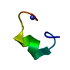
| ||||||||
|---|---|---|---|---|---|---|---|---|---|
| 1 |
| ||||||||
| Unit cell |
|
-Components
| #1: Protein/peptide | Mass: 1625.849 Da / Num. of mol.: 1 Source method: isolated from a genetically manipulated source Source: (gene. exp.) Conus pennaceus (invertebrata) / References: UniProt: P50984 |
|---|---|
| #2: Water | ChemComp-HOH /  Mass: 18.015 Da / Num. of mol.: 12 / Source method: isolated from a natural source / Formula: H2O Mass: 18.015 Da / Num. of mol.: 12 / Source method: isolated from a natural source / Formula: H2O |
| Has protein modification | Y |
-Experimental details
-Experiment
| Experiment | Method: X-RAY DIFFRACTION / Number of used crystals: 2 |
|---|
- Sample preparation
| Crystal | Density % sol: 12 % | ||||||||||||||||||||||||||||||
|---|---|---|---|---|---|---|---|---|---|---|---|---|---|---|---|---|---|---|---|---|---|---|---|---|---|---|---|---|---|---|---|
| Crystal grow | pH: 8 / Details: pH 8.0 | ||||||||||||||||||||||||||||||
| Crystal grow | *PLUS Temperature: 14 ℃ / Method: vapor diffusion, hanging drop / Details: used to seeding | ||||||||||||||||||||||||||||||
| Components of the solutions | *PLUS
|
-Data collection
| Diffraction | Mean temperature: 289 K |
|---|---|
| Diffraction source | Wavelength: 1.5418 |
| Detector | Type: RIGAKU RAXIS IIC / Detector: IMAGE PLATE / Date: Sep 28, 1995 |
| Radiation | Monochromator: YALE MIRRORS / Monochromatic (M) / Laue (L): M / Scattering type: x-ray |
| Radiation wavelength | Wavelength: 1.5418 Å / Relative weight: 1 |
| Reflection | Resolution: 1.1→50 Å / Num. obs: 3459 / % possible obs: 94 % / Observed criterion σ(I): 1 / Redundancy: 4.7 % / Rmerge(I) obs: 0.08 |
| Reflection | *PLUS Num. measured all: 16298 / Rmerge(I) obs: 0.08 |
| Reflection shell | *PLUS Highest resolution: 1.1 Å / Lowest resolution: 1.14 Å / % possible obs: 80 % / Rmerge(I) obs: 0.24 |
- Processing
| Software |
| ||||||||||||||||||||||||||||||||||||||||||||||||||||||||||||||||||||||||||||||||
|---|---|---|---|---|---|---|---|---|---|---|---|---|---|---|---|---|---|---|---|---|---|---|---|---|---|---|---|---|---|---|---|---|---|---|---|---|---|---|---|---|---|---|---|---|---|---|---|---|---|---|---|---|---|---|---|---|---|---|---|---|---|---|---|---|---|---|---|---|---|---|---|---|---|---|---|---|---|---|---|---|---|
| Refinement | Resolution: 1.1→6.1 Å / σ(F): 2
| ||||||||||||||||||||||||||||||||||||||||||||||||||||||||||||||||||||||||||||||||
| Displacement parameters | Biso mean: 7.6 Å2 | ||||||||||||||||||||||||||||||||||||||||||||||||||||||||||||||||||||||||||||||||
| Refine analyze | Luzzati coordinate error obs: 0.1 Å | ||||||||||||||||||||||||||||||||||||||||||||||||||||||||||||||||||||||||||||||||
| Refinement step | Cycle: LAST / Resolution: 1.1→6.1 Å
| ||||||||||||||||||||||||||||||||||||||||||||||||||||||||||||||||||||||||||||||||
| Refine LS restraints |
| ||||||||||||||||||||||||||||||||||||||||||||||||||||||||||||||||||||||||||||||||
| Software | *PLUS Name: X-PLOR / Version: 3.1 / Classification: refinement | ||||||||||||||||||||||||||||||||||||||||||||||||||||||||||||||||||||||||||||||||
| Refine LS restraints | *PLUS
|
