 Movie
Movie Controller

[English] 日本語
 Yorodumi
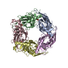
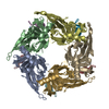
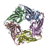
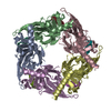
- PDB-5jme: Crystal structure of acetylcholine binding protein (AChBP) from A... -
+ Open data

- Basic information
| Entry | Database: PDB / ID: 5jme | ||||||
|---|---|---|---|---|---|---|---|
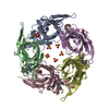
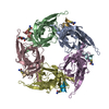
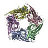
| Title | Crystal structure of acetylcholine binding protein (AChBP) from Aplysia Californica in complex with alpha-conotoxin PeIA | ||||||
 Components Components |
| ||||||
 Keywords Keywords | ACETYLCHOLINE BINDING PROTEIN / AChBP / Conotoxin / Nicotinic | ||||||
| Function / homology |  Function and homology information Function and homology informationhost cell postsynaptic membrane / acetylcholine receptor inhibitor activity / extracellular ligand-gated monoatomic ion channel activity / calcium channel regulator activity / transmembrane signaling receptor activity / toxin activity / extracellular region / membrane / metal ion binding / identical protein binding Similarity search - Function | ||||||
| Biological species |  Conus pergrandis (invertebrata) Conus pergrandis (invertebrata) | ||||||
| Method |  X-RAY DIFFRACTION / SYNCHROTRON / MOLECULAR REPLACEMENT / Resolution: 2.336 Å X-RAY DIFFRACTION / SYNCHROTRON / MOLECULAR REPLACEMENT / Resolution: 2.336 Å | ||||||
 Authors Authors | Bobango, J. / Sankaran, B. / McIntosh, J.M. / Talley, T.T. | ||||||
 Citation Citation | Journal: To Be Published Title: Crystal structure of acetylcholine binding protein (AChBP) from Aplysia Californica in complex with alpha-conotoxin PeIA Authors: Bobango, J. / McIntosh, J.M. / Sankaran, B. / Talley, T.T. | ||||||
| History |
|
- Structure visualization
| Structure viewer | Molecule: MolmilJmol/JSmol |
|---|
- Downloads & links
-Download
| PDBx/mmCIF format | 5jme.cif.gz | 227.7 KB | Display | PDBx/mmCIF format |
|---|---|---|---|---|
| PDB format | pdb5jme.ent.gz | 180.9 KB | Display | PDB format |
| PDBx/mmJSON format | 5jme.json.gz | Tree view | PDBx/mmJSON format | |
| Others |  Other downloads Other downloads |
-Validation report
| Arichive directory | https://data.pdbj.org/pub/pdb/validation_reports/jm/5jmeftp://data.pdbj.org/pub/pdb/validation_reports/jm/5jme | HTTPS FTP |
|---|
-Related structure data
| Related structure data |  4ez1S S: Starting model for refinement |
|---|---|
| Similar structure data |


-Links
 PDBj
PDBj

- Assembly
| Deposited unit | 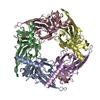
| ||||||||||
|---|---|---|---|---|---|---|---|---|---|---|---|
| 1 |
| ||||||||||
| Unit cell |
|
-Components
| #1: Protein | Mass: 26212.105 Da / Num. of mol.: 5 / Fragment: UNP residues 18-236 Source method: isolated from a genetically manipulated source Source: (gene. exp.) Production host:  Homo sapiens (human) / References: UniProt: Q8WSF8 Homo sapiens (human) / References: UniProt: Q8WSF8#2: Protein/peptide | Mass: 1656.908 Da / Num. of mol.: 4 / Source method: obtained synthetically / Details: C-terminal is aminated / Source: (synth.) Conus pergrandis (invertebrata) / References: UniProt: Q1L777*PLUS#3: Water | ChemComp-HOH / |  Mass: 18.015 Da / Num. of mol.: 332 / Source method: isolated from a natural source / Formula: H2O Mass: 18.015 Da / Num. of mol.: 332 / Source method: isolated from a natural source / Formula: H2OHas protein modification | Y | |
|---|
-Experimental details
-Experiment
| Experiment | Method: X-RAY DIFFRACTION / Number of used crystals: 1 |
|---|
- Sample preparation
| Crystal | Density Matthews: 2.85 Å3/Da / Density % sol: 61.18 % |
|---|---|
| Crystal grow | Temperature: 290 K / Method: vapor diffusion, hanging drop / pH: 8 Details: 0.1 M Tris-HCl, 0.25 M magnesium chloride, 20% w/v PEG4000 |
-Data collection
| Diffraction | Mean temperature: 200 K |
|---|---|
| Diffraction source | Source: SYNCHROTRON / Site: ALS  / Beamline: 8.2.1 / Wavelength: 1 Å / Beamline: 8.2.1 / Wavelength: 1 Å |
| Detector | Type: ADSC QUANTUM 315r / Detector: CCD / Date: Oct 5, 2007 |
| Radiation | Protocol: SINGLE WAVELENGTH / Monochromatic (M) / Laue (L): M / Scattering type: x-ray |
| Radiation wavelength | Wavelength: 1 Å / Relative weight: 1 |
| Reflection | Resolution: 2.336→48.821 Å / Num. obs: 66090 / % possible obs: 92.6 % / Redundancy: 14.75 % / Biso Wilson estimate: 36.0398620104 Å2 / Net I/σ(I): 3.51 |
- Processing
| Software |
| |||||||||||||||||||||||||||||||||||||||||||||||||||||||||||||||||||||||||||||||||||||||||||||||||||||||||
|---|---|---|---|---|---|---|---|---|---|---|---|---|---|---|---|---|---|---|---|---|---|---|---|---|---|---|---|---|---|---|---|---|---|---|---|---|---|---|---|---|---|---|---|---|---|---|---|---|---|---|---|---|---|---|---|---|---|---|---|---|---|---|---|---|---|---|---|---|---|---|---|---|---|---|---|---|---|---|---|---|---|---|---|---|---|---|---|---|---|---|---|---|---|---|---|---|---|---|---|---|---|---|---|---|---|---|
| Refinement | Method to determine structure: MOLECULAR REPLACEMENT Starting model: 4EZ1 Resolution: 2.336→48.821 Å / SU ML: 0.25 / Cross valid method: NONE / σ(F): 1.34 / Phase error: 25.13 / Stereochemistry target values: ML
| |||||||||||||||||||||||||||||||||||||||||||||||||||||||||||||||||||||||||||||||||||||||||||||||||||||||||
| Solvent computation | Shrinkage radii: 0.9 Å / VDW probe radii: 1.11 Å / Solvent model: FLAT BULK SOLVENT MODEL | |||||||||||||||||||||||||||||||||||||||||||||||||||||||||||||||||||||||||||||||||||||||||||||||||||||||||
| Displacement parameters | Biso max: 99.75 Å2 / Biso mean: 39.913 Å2 / Biso min: 18.69 Å2 | |||||||||||||||||||||||||||||||||||||||||||||||||||||||||||||||||||||||||||||||||||||||||||||||||||||||||
| Refinement step | Cycle: final / Resolution: 2.336→48.821 Å
| |||||||||||||||||||||||||||||||||||||||||||||||||||||||||||||||||||||||||||||||||||||||||||||||||||||||||
| Refine LS restraints |
| |||||||||||||||||||||||||||||||||||||||||||||||||||||||||||||||||||||||||||||||||||||||||||||||||||||||||
| LS refinement shell | Refine-ID: X-RAY DIFFRACTION / Total num. of bins used: 14
|
