 Movie
Movie Controller

[English] 日本語
 Yorodumi
- PDB-4zjt: X-ray crystal structure of Lymnaea stagnalis acetylcholine bindin... -
+ Open data

- Basic information
| Entry | Database: PDB / ID: 4zjt | ||||||
|---|---|---|---|---|---|---|---|
| Title | X-ray crystal structure of Lymnaea stagnalis acetylcholine binding protein (LsAChBP) in complex with 2-Thiophenylmethylene Anabaseine (2TAB) | ||||||
 Components Components | Acetylcholine-binding protein | ||||||
 Keywords Keywords | ACETYLCHOLINE BINDING PROTEIN / nicotinic / AChBP / receptor / Anabaseine | ||||||
| Function / homology |  Function and homology information Function and homology informationsynaptic cleft / extracellular ligand-gated monoatomic ion channel activity / transmembrane signaling receptor activity / membrane Similarity search - Function | ||||||
| Biological species |   Lymnaea stagnalis (great pond snail) Lymnaea stagnalis (great pond snail) | ||||||
| Method |  X-RAY DIFFRACTION / SYNCHROTRON / MOLECULAR REPLACEMENT / Resolution: 1.85 Å X-RAY DIFFRACTION / SYNCHROTRON / MOLECULAR REPLACEMENT / Resolution: 1.85 Å | ||||||
 Authors Authors | Bobango, J. / Wu, J. / Talley, T.T. | ||||||
 Citation Citation | Journal: To Be Published Title: X-ray crystal structure of Lymnaea stagnalis acetylcholine binding protein (LsAChBP) in complex with 2-Thiophenylmethylene Anabaseine (2TAB) Authors: Bobango, J. / Wu, J. / Talley, T.T. | ||||||
| History |
|
- Structure visualization
| Structure viewer | Molecule: MolmilJmol/JSmol |
|---|
- Downloads & links
-Download
| PDBx/mmCIF format | 4zjt.cif.gz | 434.3 KB | Display | PDBx/mmCIF format |
|---|---|---|---|---|
| PDB format | pdb4zjt.ent.gz | 353.2 KB | Display | PDB format |
| PDBx/mmJSON format | 4zjt.json.gz | Tree view | PDBx/mmJSON format | |
| Others |  Other downloads Other downloads |
-Validation report
| Arichive directory | https://data.pdbj.org/pub/pdb/validation_reports/zj/4zjtftp://data.pdbj.org/pub/pdb/validation_reports/zj/4zjt | HTTPS FTP |
|---|
-Related structure data
| Related structure data |  1uv6S  1uw6S 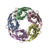 1ux2S 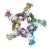 1yi5S  2zjuS  2zjvS S: Starting model for refinement |
|---|---|
| Similar structure data |


-Links
 PDBj
PDBj

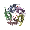
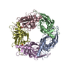
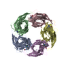
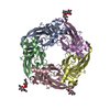
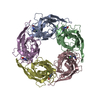
- Assembly
| Deposited unit | 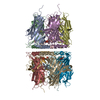
| ||||||||
|---|---|---|---|---|---|---|---|---|---|
| 1 | 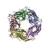
| ||||||||
| 2 | 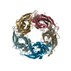
| ||||||||
| Unit cell |
|
-Components
| #1: Protein | Mass: 24859.496 Da / Num. of mol.: 10 / Fragment: UNP residues 20-229 Source method: isolated from a genetically manipulated source Source: (gene. exp.) Lymnaea stagnalis (great pond snail) / Cell line (production host): HEK293S GnTI- / Production host:  Homo sapiens (human) / References: UniProt: P58154 Homo sapiens (human) / References: UniProt: P58154#2: Chemical | ChemComp-PO4 /   Mass: 94.971 Da / Num. of mol.: 10 / Source method: obtained synthetically / Formula: PO4 Mass: 94.971 Da / Num. of mol.: 10 / Source method: obtained synthetically / Formula: PO4#3: Chemical | ChemComp-4P6 / ( 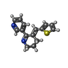  Mass: 254.350 Da / Num. of mol.: 10 / Source method: obtained synthetically / Formula: C15H14N2S Mass: 254.350 Da / Num. of mol.: 10 / Source method: obtained synthetically / Formula: C15H14N2S#4: Water | ChemComp-HOH / |  Mass: 18.015 Da / Num. of mol.: 656 / Source method: isolated from a natural source / Formula: H2O Mass: 18.015 Da / Num. of mol.: 656 / Source method: isolated from a natural source / Formula: H2OHas protein modification | Y | |
|---|
-Experimental details
-Experiment
| Experiment | Method: X-RAY DIFFRACTION / Number of used crystals: 1 |
|---|
- Sample preparation
| Crystal | Density Matthews: 2.53 Å3/Da / Density % sol: 51.35 % |
|---|---|
| Crystal grow | Temperature: 293 K / Method: vapor diffusion, hanging drop / Details: 0.26 M ammonium phosphate, 35% glycerol |
-Data collection
| Diffraction | Mean temperature: 88 K |
|---|---|
| Diffraction source | Source: SYNCHROTRON / Site: ALS  / Beamline: 8.2.1 / Wavelength: 1 Å / Beamline: 8.2.1 / Wavelength: 1 Å |
| Detector | Type: ADSC QUANTUM 315r / Detector: CCD / Date: Dec 20, 2011 |
| Diffraction measurement | Details: 1.00 degrees, 1.0 sec, detector distance 250.00 mm |
| Radiation | Monochromator: double crystal Si(111) / Protocol: SINGLE WAVELENGTH / Monochromatic (M) / Laue (L): M / Scattering type: x-ray |
| Radiation wavelength | Wavelength: 1 Å / Relative weight: 1 |
| Reflection | Av R equivalents: 0.077 / Number: 1708039 |
| Reflection | Resolution: 1.74→50 Å / Num. obs: 248133 / % possible obs: 96 % / Observed criterion σ(F): 0 / Observed criterion σ(I): -3 / Redundancy: 7.1 % / Rmerge(I) obs: 0.077 / Rsym value: 0.077 / Net I/av σ(I): 22.167 / Net I/σ(I): 23.8 |
| Reflection shell | Resolution: 1.74→1.77 Å / Redundancy: 4.4 % / Rmerge(I) obs: 0.035 / Mean I/σ(I) obs: 1.237 / Rsym value: 0.688 / % possible all: 68.5 |
| Cell measurement | Reflection used: 1708039 |
- Processing
| Software |
| |||||||||||||||||||||||||||||||||||||||||||||||||||||||||||||||||||||||||||||||||||||||||||
|---|---|---|---|---|---|---|---|---|---|---|---|---|---|---|---|---|---|---|---|---|---|---|---|---|---|---|---|---|---|---|---|---|---|---|---|---|---|---|---|---|---|---|---|---|---|---|---|---|---|---|---|---|---|---|---|---|---|---|---|---|---|---|---|---|---|---|---|---|---|---|---|---|---|---|---|---|---|---|---|---|---|---|---|---|---|---|---|---|---|---|---|---|
| Refinement | Method to determine structure: MOLECULAR REPLACEMENT Starting model: PDB entries 1YI5, 1UV6, 1UW6, 1UX2, 2ZJU, 2ZJV Resolution: 1.85→49.985 Å / SU ML: 0.21 / σ(F): 1.34 / Phase error: 24.57 / Stereochemistry target values: ML
| |||||||||||||||||||||||||||||||||||||||||||||||||||||||||||||||||||||||||||||||||||||||||||
| Solvent computation | Shrinkage radii: 0.9 Å / VDW probe radii: 1.11 Å / Solvent model: FLAT BULK SOLVENT MODEL | |||||||||||||||||||||||||||||||||||||||||||||||||||||||||||||||||||||||||||||||||||||||||||
| Displacement parameters | Biso max: 77.68 Å2 / Biso mean: 26.4473 Å2 / Biso min: 12.21 Å2 | |||||||||||||||||||||||||||||||||||||||||||||||||||||||||||||||||||||||||||||||||||||||||||
| Refinement step | Cycle: final / Resolution: 1.85→49.985 Å
| |||||||||||||||||||||||||||||||||||||||||||||||||||||||||||||||||||||||||||||||||||||||||||
| Refine LS restraints |
| |||||||||||||||||||||||||||||||||||||||||||||||||||||||||||||||||||||||||||||||||||||||||||
| LS refinement shell | Refine-ID: X-RAY DIFFRACTION / Total num. of bins used: 12
|
