Mass: 18.015 Da / Num. of mol.: 399 / Source method: isolated from a natural source / Formula: H2O
Has protein modification
Y
Sequence details
THE AUTHORS STATE THAT THE SEQUENCE BETWEEN RESIDUES 69 AND 93 IN THE DBREF RECORDS BELOW REPRESENT ...THE AUTHORS STATE THAT THE SEQUENCE BETWEEN RESIDUES 69 AND 93 IN THE DBREF RECORDS BELOW REPRESENT THE TRUE SEQUENCE OF THE PROTEIN. THE CORRESPONDING UNIPROT CONTAINS THE RESULTS OF A SEQUENCING ERROR DISCOVERED DURING THE PREVIOUS DETERMINATION OF THE STRUCTURE (PDB ENTRY 1H0B).
-
Experimental details
-
Experiment
Experiment
Method: X-RAY DIFFRACTION / Number of used crystals: 1
-
Sample preparation
Crystal
Density Matthews: 2.5 Å3/Da / Density % sol: 41.5 %
Crystal grow
pH: 6.5 Details: 0.1M MES PH 6.5, 1.5M (NH4)2SO4, 0.2M LI2SO4, 10MG/ML PROTEIN
In the structure databanks used in Yorodumi, some data are registered as the other names, "COVID-19 virus" and "2019-nCoV". Here are the details of the virus and the list of structure data.
Jan 31, 2019. EMDB accession codes are about to change! (news from PDBe EMDB page)
EMDB accession codes are about to change! (news from PDBe EMDB page)
The allocation of 4 digits for EMDB accession codes will soon come to an end. Whilst these codes will remain in use, new EMDB accession codes will include an additional digit and will expand incrementally as the available range of codes is exhausted. The current 4-digit format prefixed with “EMD-” (i.e. EMD-XXXX) will advance to a 5-digit format (i.e. EMD-XXXXX), and so on. It is currently estimated that the 4-digit codes will be depleted around Spring 2019, at which point the 5-digit format will come into force.
The EM Navigator/Yorodumi systems omit the EMD- prefix.
Related info.:Q: What is EMD? / ID/Accession-code notation in Yorodumi/EM Navigator
Yorodumi is a browser for structure data from EMDB, PDB, SASBDB, etc.
This page is also the successor to EM Navigator detail page, and also detail information page/front-end page for Omokage search.
The word "yorodu" (or yorozu) is an old Japanese word meaning "ten thousand". "mi" (miru) is to see.
Related info.:EMDB / PDB / SASBDB / Comparison of 3 databanks / Yorodumi Search / Aug 31, 2016. New EM Navigator & Yorodumi / Yorodumi Papers / Jmol/JSmol / Function and homology information / Changes in new EM Navigator and Yorodumi
 Movie
Movie Controller

 Yorodumi
 Open data
 Basic information
 Components
 Keywords
 Function and homology information
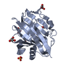
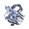
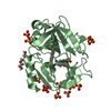
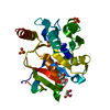
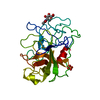
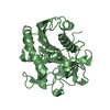
 RHODOTHERMUS MARINUS (bacteria)
 X-RAY DIFFRACTION /
  Authors
 Citation
 Structure visualization
 Downloads & links
 Other downloads
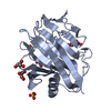


 PDBj
PDBj


 Assembly
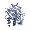


 Mass: 96.063 Da / Num. of mol.: 1 / Source method: obtained synthetically / Formula: SO4
 Mass: 18.015 Da / Num. of mol.: 399 / Source method: isolated from a natural source / Formula: H2O
 Sample preparation
 / Beamline: BM14 / Wavelength: 0.97845
  Processing