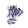
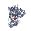
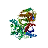
[English] 日本語
Yorodumi
- PDB-2bra: Structure of N-Terminal FAD Binding motif of mouse MICAL -

+
Open data


ID or keywords:

Loading...

-
Basic information

Entry
Database: PDB / ID: 2bra
TitleStructure of N-Terminal FAD Binding motif of mouse MICAL
ComponentsNEDD9 INTERACTING PROTEIN WITH CALPONIN HOMOLOGY AND LIM DOMAINS
KeywordsTRANSPORT / AXON GUIDANCE / VESICLE TRANSPORT / FLAVOPROTEIN / REDOX / PLEXIN / COILED COIL / CYTOSKELETON / FAD / LIM DOMAIN / METAL-BINDING / ZINC
Function / homology
Function and homology information


hippocampal mossy fiber expansion / NADPH oxidase H202-forming activity / F-actin monooxygenase / NAD(P)H oxidase (H2O2-forming) / sulfur oxidation / regulation of regulated secretory pathway / oxidoreductase activity, acting on paired donors, with incorporation or reduction of molecular oxygen, NAD(P)H as one donor, and incorporation of one atom of oxygen / actin filament depolymerization / intercellular bridge / monooxygenase activity ...hippocampal mossy fiber expansion / NADPH oxidase H202-forming activity / F-actin monooxygenase / NAD(P)H oxidase (H2O2-forming) / sulfur oxidation / regulation of regulated secretory pathway / oxidoreductase activity, acting on paired donors, with incorporation or reduction of molecular oxygen, NAD(P)H as one donor, and incorporation of one atom of oxygen / actin filament depolymerization / intercellular bridge / monooxygenase activity / FAD binding / negative regulation of protein phosphorylation / actin filament / negative regulation of cysteine-type endopeptidase activity involved in apoptotic process / small GTPase binding / SH3 domain binding / actin binding / midbody / endosome membrane / negative regulation of apoptotic process / protein kinase binding / metal ion binding / cytoplasm / cytosol
Similarity search - Function
DUF3585 / bMERB domain / Bivalent Mical/EHBP Rab binding domain / bMERB domain profile. / LIM zinc-binding domain signature. / Zinc-binding domain present in Lin-11, Isl-1, Mec-3. / Zinc finger, LIM-type / LIM domain profile. / FAD-binding domain / FAD binding domain ...DUF3585 / bMERB domain / Bivalent Mical/EHBP Rab binding domain / bMERB domain profile. / LIM zinc-binding domain signature. / Zinc-binding domain present in Lin-11, Isl-1, Mec-3. / Zinc finger, LIM-type / LIM domain profile. / FAD-binding domain / FAD binding domain / Calponin homology domain / Calponin homology (CH) domain / Calponin homology domain / CH domain superfamily / Calponin homology (CH) domain profile. / FAD/NAD(P)-binding domain / FAD/NAD(P)-binding domain / 3-Layer(bba) Sandwich / FAD/NAD(P)-binding domain superfamily / Alpha Beta
Similarity search - Domain/homology
FLAVIN-ADENINE DINUCLEOTIDE / [F-actin]-monooxygenase MICAL1
Similarity search - Component
Biological speciesMUS MUSCULUS (house mouse)
MethodX-RAY DIFFRACTION / SYNCHROTRON / SIRAS / Resolution: 2 Å
AuthorsNadella, M. / Bianchet, M.A. / Gabelli, S.B. / Amzel, L.M.
CitationJournal: Proc. Natl. Acad. Sci. U.S.A. / Year: 2005
Title: Structure and activity of the axon guidance protein MICAL.
Authors: Nadella, M. / Bianchet, M.A. / Gabelli, S.B. / Barrila, J. / Amzel, L.M.
History
DepositionMay 4, 2005Deposition site: PDBE / Processing site: PDBE
Revision 1.0Nov 1, 2005Provider: repository / Type: Initial release
Revision 1.1May 8, 2011Group: Version format compliance
Revision 1.2Jul 13, 2011Group: Version format compliance
Revision 1.3Feb 28, 2018Group: Database references / Source and taxonomy / Category: citation / entity_src_gen
Item: _citation.journal_abbrev / _citation.page_last ..._citation.journal_abbrev / _citation.page_last / _citation.pdbx_database_id_DOI / _citation.title / _entity_src_gen.pdbx_host_org_ncbi_taxonomy_id / _entity_src_gen.pdbx_host_org_scientific_name / _entity_src_gen.pdbx_host_org_strain / _entity_src_gen.pdbx_host_org_variant
Revision 1.4Mar 14, 2018Group: Source and taxonomy / Category: entity_src_gen / Item: _entity_src_gen.pdbx_host_org_cell_line
Revision 1.5May 8, 2024Group: Data collection / Database references / Category: chem_comp_atom / chem_comp_bond / database_2
Item: _database_2.pdbx_DOI / _database_2.pdbx_database_accession

-
Structure visualization

Structure viewerMolecule:
MolmilJmol/JSmol

Downloads & links

-
Assembly

Deposited unit
A: NEDD9 INTERACTING PROTEIN WITH CALPONIN HOMOLOGY AND LIM DOMAINS
B: NEDD9 INTERACTING PROTEIN WITH CALPONIN HOMOLOGY AND LIM DOMAINS
hetero molecules


Theoretical massNumber of molelcules
Total (without water)108,5155
Polymers106,9092
Non-polymers1,6073
Water11,115617
1
A: NEDD9 INTERACTING PROTEIN WITH CALPONIN HOMOLOGY AND LIM DOMAINS
hetero molecules


Theoretical massNumber of molelcules
Total (without water)54,2402
Polymers53,4541
Non-polymers7861
Water181
TypeNameSymmetry operationNumber
identity operation1_555x,y,z1
MethodPQS
2
B: NEDD9 INTERACTING PROTEIN WITH CALPONIN HOMOLOGY AND LIM DOMAINS
hetero molecules


Theoretical massNumber of molelcules
Total (without water)54,2753
Polymers53,4541
Non-polymers8212
Water181
TypeNameSymmetry operationNumber
identity operation1_555x,y,z1
MethodPQS
Unit cell
Length a, b, c (Å)72.969, 87.338, 80.810
Angle α, β, γ (deg.)90.00, 111.68, 90.00
Int Tables number4
Space group name H-MP1211

-
Components

#1: Protein NEDD9 INTERACTING PROTEIN WITH CALPONIN HOMOLOGY AND LIM DOMAINS / MICAL / MOLECULE INTERACTING WITH CASL PROTEIN 1


Mass: 53454.297 Da / Num. of mol.: 2 / Fragment: N-TERMINAL DOMAIN, RESIDUES 1-484 / Mutation: YES
Source method: isolated from a genetically manipulated source
Source: (gene. exp.) MUS MUSCULUS (house mouse) / Tissue: MAMMARY TUMOR / Production host: Escherichia coli BL21(DE3) (bacteria) / Variant (production host): Rosetta pLysS / References: UniProt: Q8VDP3
#2: Chemical ChemComp-FAD / FLAVIN-ADENINE DINUCLEOTIDE


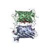
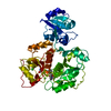
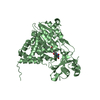
Mass: 785.550 Da / Num. of mol.: 2 / Source method: obtained synthetically / Formula: C27H33N9O15P2 / Comment: FAD*YM
#3: Chemical ChemComp-CL / CHLORIDE ION


Mass: 35.453 Da / Num. of mol.: 1 / Source method: obtained synthetically / Formula: Cl
#4: Water ChemComp-HOH / water


Mass: 18.015 Da / Num. of mol.: 617 / Source method: isolated from a natural source / Formula: H2O
Compound detailsPOSSIBLE CYTOSKELETAL REGULATOR THAT CONNECTS NEDD9 TO INTERMEDIATE FILAMENTS ENGINEERED RESIDUE IN ...POSSIBLE CYTOSKELETAL REGULATOR THAT CONNECTS NEDD9 TO INTERMEDIATE FILAMENTS ENGINEERED RESIDUE IN CHAIN A, LYS 141 TO ALA ENGINEERED RESIDUE IN CHAIN A, LYS 142 TO ALA ENGINEERED RESIDUE IN CHAIN B, LYS 141 TO ALA ENGINEERED RESIDUE IN CHAIN B, LYS 142 TO ALA

-
Experimental details

-
Experiment

ExperimentMethod: X-RAY DIFFRACTION / Number of used crystals: 1

-
Sample preparation

CrystalDensity Matthews: 2.2 Å3/Da / Density % sol: 47 %
Crystal growpH: 4.6
Details: PROTEIN SAMPLE: 20 MM HEPES PH 7.0, 1M NACL, 1MM DTT, 4 MG/ML MICAL RECERVOIR: 100MM NA ACETATE PH 4.6, 42% W/V PEG 2K MME 0.3 M (NH4)2SO4

-
Data collection

DiffractionMean temperature: 100 K
Diffraction sourceSource: SYNCHROTRON / Site: NSLS / Beamline: X6A / Wavelength: 1.1
DetectorType: ADSC CCD / Detector: CCD / Date: Feb 14, 2005 / Details: MIRRORS
RadiationProtocol: SINGLE WAVELENGTH / Monochromatic (M) / Laue (L): M / Scattering type: x-ray
Radiation wavelengthWavelength: 1.1 Å / Relative weight: 1
ReflectionResolution: 2→50 Å / Num. obs: 62811 / % possible obs: 98.9 % / Observed criterion σ(I): 0 / Redundancy: 3.5 % / Rmerge(I) obs: 0.11 / Net I/σ(I): 15.2
Reflection shellResolution: 2→2.07 Å / Redundancy: 2.7 % / Rmerge(I) obs: 0.59 / Mean I/σ(I) obs: 1.56 / % possible all: 94

-
Processing

Software
NameVersionClassification
REFMAC5.2.0005refinement
HKL-2000data reduction
SCALEPACKdata scaling
SOLVEphasing
RefinementMethod to determine structure: SIRAS / Resolution: 2→27.94 Å / Cor.coef. Fo:Fc: 0.959 / Cor.coef. Fo:Fc free: 0.92 / SU B: 11.6 / SU ML: 0.165 / Cross valid method: THROUGHOUT / ESU R: 0.213 / ESU R Free: 0.2 / Stereochemistry target values: MAXIMUM LIKELIHOOD / Details: HYDROGENS HAVE BEEN ADDED IN THE RIDING POSITIONS.
RfactorNum. reflection% reflectionSelection details
Rfree0.266 3124 5 %RANDOM
Rwork0.192 ---
obs0.195 59660 100 %-
Solvent computationIon probe radii: 0.8 Å / Shrinkage radii: 0.8 Å / VDW probe radii: 1.2 Å / Solvent model: MASK
Displacement parametersBiso mean: 35.55 Å2
Baniso -1Baniso -2Baniso -3
1--1.58 Å20 Å2-2 Å2
2--2.33 Å20 Å2
3----2.23 Å2
Refinement stepCycle: LAST / Resolution: 2→27.94 Å
ProteinNucleic acidLigandSolventTotal
Num. atoms7413 0 107 617 8137
Refine LS restraints
Refine-IDTypeDev idealDev ideal targetNumber
X-RAY DIFFRACTIONr_bond_refined_d0.020.0227692
X-RAY DIFFRACTIONr_bond_other_d
X-RAY DIFFRACTIONr_angle_refined_deg1.9761.97410441
X-RAY DIFFRACTIONr_angle_other_deg
X-RAY DIFFRACTIONr_dihedral_angle_1_deg7.2515946
X-RAY DIFFRACTIONr_dihedral_angle_2_deg39.41223.362345
X-RAY DIFFRACTIONr_dihedral_angle_3_deg19.778151264
X-RAY DIFFRACTIONr_dihedral_angle_4_deg20.6241560
X-RAY DIFFRACTIONr_chiral_restr0.1460.21147
X-RAY DIFFRACTIONr_gen_planes_refined0.0080.025822
X-RAY DIFFRACTIONr_gen_planes_other
X-RAY DIFFRACTIONr_nbd_refined0.2460.23911
X-RAY DIFFRACTIONr_nbd_other
X-RAY DIFFRACTIONr_nbtor_refined0.3210.25181
X-RAY DIFFRACTIONr_nbtor_other
X-RAY DIFFRACTIONr_xyhbond_nbd_refined0.1860.2648
X-RAY DIFFRACTIONr_xyhbond_nbd_other
X-RAY DIFFRACTIONr_metal_ion_refined
X-RAY DIFFRACTIONr_metal_ion_other
X-RAY DIFFRACTIONr_symmetry_vdw_refined0.2950.2121
X-RAY DIFFRACTIONr_symmetry_vdw_other
X-RAY DIFFRACTIONr_symmetry_hbond_refined0.2330.224
X-RAY DIFFRACTIONr_symmetry_hbond_other
X-RAY DIFFRACTIONr_symmetry_metal_ion_refined
X-RAY DIFFRACTIONr_symmetry_metal_ion_other
X-RAY DIFFRACTIONr_mcbond_it1.1891.54830
X-RAY DIFFRACTIONr_mcbond_other
X-RAY DIFFRACTIONr_mcangle_it1.86627518
X-RAY DIFFRACTIONr_mcangle_other
X-RAY DIFFRACTIONr_scbond_it2.84633326
X-RAY DIFFRACTIONr_scbond_other
X-RAY DIFFRACTIONr_scangle_it4.3514.52923
X-RAY DIFFRACTIONr_scangle_other
X-RAY DIFFRACTIONr_long_range_B_refined
X-RAY DIFFRACTIONr_long_range_B_other
X-RAY DIFFRACTIONr_rigid_bond_restr
X-RAY DIFFRACTIONr_sphericity_free
X-RAY DIFFRACTIONr_sphericity_bonded
LS refinement shellResolution: 2→2.05 Å / Total num. of bins used: 20 /
RfactorNum. reflection
Rfree0.354 220
Rwork0.253 3836
Refinement TLS params.

Method: refined / Refine-ID: X-RAY DIFFRACTION

IDL112)L122)L132)L222)L232)L332)S11 (Å °)S12 (Å °)S13 (Å °)S21 (Å °)S22 (Å °)S23 (Å °)S31 (Å °)S32 (Å °)S33 (Å °)T112)T122)T132)T222)T232)T332)Origin x (Å)Origin y (Å)Origin z (Å)
10.69880.05920.06820.6103-0.28950.55330.03170.0523-0.0401-0.01450.0110.0120.04690.0354-0.04280.0353-0.0061-0.0056-0.1135-0.01640.021741.6126.904-6.317
22.0290.1537-1.32611.0766-0.0885.89780.0302-0.1126-0.09110.0801-0.0509-0.12860.05540.39040.0208-0.03070.00160.0052-0.10030.0004-0.003168.57141.0453.245
30.7872-0.02360.54940.5020.08091.81910.0348-0.0194-0.0578-0.01870.0189-0.01670.082-0.1045-0.0537-0.00060.01770.0004-0.10150.0134-0.001854.08724.26441.71
41.84060.0934-1.10930.7158-0.427510.13870.06850.0968-0.0266-0.07630.02590.1471-0.1003-0.1905-0.0945-0.03770.0304-0.0043-0.11250.008-0.032330.04543.27232.875
Refinement TLS group
IDRefine-IDRefine TLS-IDAuth asym-IDAuth seq-ID
1X-RAY DIFFRACTION1A8 - 234
2X-RAY DIFFRACTION1A368 - 520
3X-RAY DIFFRACTION2A235 - 369
4X-RAY DIFFRACTION3B1 - 234
5X-RAY DIFFRACTION3B368 - 520
6X-RAY DIFFRACTION4B235 - 367

+
About Yorodumi

-
News

-
Feb 9, 2022. New format data for meta-information of EMDB entries

New format data for meta-information of EMDB entries

  • Version 3 of the EMDB header file is now the official format.
  • The previous official version 1.9 will be removed from the archive.

Related info.:EMDB header

External links:wwPDB to switch to version 3 of the EMDB data model

-
Aug 12, 2020. Covid-19 info

Covid-19 info

URL: https://pdbjlvh1.pdbj.org/emnavi/covid19.php

New page: Covid-19 featured information page in EM Navigator.

Related info.:Covid-19 info / Mar 5, 2020. Novel coronavirus structure data

+
Mar 5, 2020. Novel coronavirus structure data

Novel coronavirus structure data

Related info.:Yorodumi Speices / Aug 12, 2020. Covid-19 info

External links:COVID-19 featured content - PDBj / Molecule of the Month (242):Coronavirus Proteases

+
Jan 31, 2019. EMDB accession codes are about to change! (news from PDBe EMDB page)

EMDB accession codes are about to change! (news from PDBe EMDB page)

  • The allocation of 4 digits for EMDB accession codes will soon come to an end. Whilst these codes will remain in use, new EMDB accession codes will include an additional digit and will expand incrementally as the available range of codes is exhausted. The current 4-digit format prefixed with “EMD-” (i.e. EMD-XXXX) will advance to a 5-digit format (i.e. EMD-XXXXX), and so on. It is currently estimated that the 4-digit codes will be depleted around Spring 2019, at which point the 5-digit format will come into force.
  • The EM Navigator/Yorodumi systems omit the EMD- prefix.

Related info.:Q: What is EMD? / ID/Accession-code notation in Yorodumi/EM Navigator

External links:EMDB Accession Codes are Changing Soon! / Contact to PDBj

+
Jul 12, 2017. Major update of PDB

Major update of PDB

  • wwPDB released updated PDB data conforming to the new PDBx/mmCIF dictionary.
  • This is a major update changing the version number from 4 to 5, and with Remediation, in which all the entries are updated.
  • In this update, many items about electron microscopy experimental information are reorganized (e.g. em_software).
  • Now, EM Navigator and Yorodumi are based on the updated data.

External links:wwPDB Remediation / Enriched Model Files Conforming to OneDep Data Standards Now Available in the PDB FTP Archive

-
Yorodumi

Thousand views of thousand structures

  • Yorodumi is a browser for structure data from EMDB, PDB, SASBDB, etc.
  • This page is also the successor to EM Navigator detail page, and also detail information page/front-end page for Omokage search.
  • The word "yorodu" (or yorozu) is an old Japanese word meaning "ten thousand". "mi" (miru) is to see.

Related info.:EMDB / PDB / SASBDB / Comparison of 3 databanks / Yorodumi Search / Aug 31, 2016. New EM Navigator & Yorodumi / Yorodumi Papers / Jmol/JSmol / Function and homology information / Changes in new EM Navigator and Yorodumi

Read more