 Movie
Movie Controller

[English] 日本語
 Yorodumi
- PDB-4q7r: Crystal structure of large Stokes shift fluorescent protein LSSmOrange -
+ Open data

- Basic information
| Entry | Database: PDB / ID: 4q7r | ||||||
|---|---|---|---|---|---|---|---|
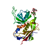
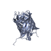
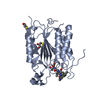
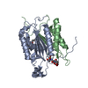
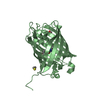
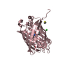
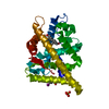
| Title | Crystal structure of large Stokes shift fluorescent protein LSSmOrange | ||||||
 Components Components | LSSmOrange | ||||||
 Keywords Keywords | FLUORESCENT PROTEIN / beta-barrel / large Stokes shift fluorescent protein / chromophore | ||||||
| Function / homology |  Function and homology information Function and homology informationbioluminescence / generation of precursor metabolites and energy / metal ion binding Similarity search - Function | ||||||
| Biological species |  Discosoma sp. (sea anemone) Discosoma sp. (sea anemone) | ||||||
| Method |  X-RAY DIFFRACTION / SYNCHROTRON / MOLECULAR REPLACEMENT / Resolution: 1.4 Å X-RAY DIFFRACTION / SYNCHROTRON / MOLECULAR REPLACEMENT / Resolution: 1.4 Å | ||||||
 Authors Authors | Pletnev, S. / Dauter, Z. | ||||||
 Citation Citation | Journal: Plos One / Year: 2014 Title: Orange Fluorescent Proteins: Structural Studies of LSSmOrange, PSmOrange and PSmOrange2. Authors: Pletnev, S. / Shcherbakova, D.M. / Subach, O.M. / Pletneva, N.V. / Malashkevich, V.N. / Almo, S.C. / Dauter, Z. / Verkhusha, V.V. | ||||||
| History |
|
- Structure visualization
| Structure viewer | Molecule: MolmilJmol/JSmol |
|---|
- Downloads & links
-Download
| PDBx/mmCIF format | 4q7r.cif.gz | 217.7 KB | Display | PDBx/mmCIF format |
|---|---|---|---|---|
| PDB format | pdb4q7r.ent.gz | 173.3 KB | Display | PDB format |
| PDBx/mmJSON format | 4q7r.json.gz | Tree view | PDBx/mmJSON format | |
| Others |  Other downloads Other downloads |
-Validation report
| Arichive directory | https://data.pdbj.org/pub/pdb/validation_reports/q7/4q7rftp://data.pdbj.org/pub/pdb/validation_reports/q7/4q7r | HTTPS FTP |
|---|
-Related structure data
| Related structure data | 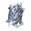 4q7tC 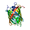 4q7uC 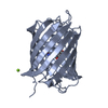 2h5oS S: Starting model for refinement C: citing same article ( |
|---|---|
| Similar structure data |

-Links
 PDBj
PDBj

- Assembly
| Deposited unit | 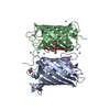
| ||||||||
|---|---|---|---|---|---|---|---|---|---|
| 1 | 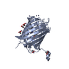
| ||||||||
| 2 | 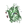
| ||||||||
| Unit cell |
|
-Components
| #1: Protein | Mass: 28120.748 Da / Num. of mol.: 2 / Mutation: R17H, V44A, F83L, W143M, I161D, M163L, G196D Source method: isolated from a genetically manipulated source Source: (gene. exp.) Discosoma sp. (sea anemone) / Production host:  #2: Chemical | ChemComp-ZN /   Mass: 65.409 Da / Num. of mol.: 17 / Source method: obtained synthetically / Formula: Zn Mass: 65.409 Da / Num. of mol.: 17 / Source method: obtained synthetically / Formula: Zn#3: Chemical | ChemComp-ACT /   Mass: 59.044 Da / Num. of mol.: 9 / Source method: obtained synthetically / Formula: C2H3O2 Mass: 59.044 Da / Num. of mol.: 9 / Source method: obtained synthetically / Formula: C2H3O2#4: Water | ChemComp-HOH / |  Mass: 18.015 Da / Num. of mol.: 301 / Source method: isolated from a natural source / Formula: H2O Mass: 18.015 Da / Num. of mol.: 301 / Source method: isolated from a natural source / Formula: H2OHas protein modification | Y | Sequence details | RESIDUES PHE65, GLY66, TYR67, AND GLY68 CIRCULARIZ | |
|---|
-Experimental details
-Experiment
| Experiment | Method: X-RAY DIFFRACTION / Number of used crystals: 1 |
|---|
- Sample preparation
| Crystal | Density Matthews: 1.98 Å3/Da / Density % sol: 37.79 % |
|---|---|
| Crystal grow | Temperature: 293 K / Method: vapor diffusion, hanging drop / pH: 4.5 Details: 0.2 M zinc acetate, 0.1 M acetate buffer, 20% PEG1000, pH 4.5, VAPOR DIFFUSION, HANGING DROP, temperature 293K |
-Data collection
| Diffraction | Mean temperature: 100 K | |||||||||||||||||||||||||||||||||||||||||||||||||||||||||||||||||||||||||||||
|---|---|---|---|---|---|---|---|---|---|---|---|---|---|---|---|---|---|---|---|---|---|---|---|---|---|---|---|---|---|---|---|---|---|---|---|---|---|---|---|---|---|---|---|---|---|---|---|---|---|---|---|---|---|---|---|---|---|---|---|---|---|---|---|---|---|---|---|---|---|---|---|---|---|---|---|---|---|---|
| Diffraction source | Source: SYNCHROTRON / Site: APS  / Beamline: 22-ID / Wavelength: 1 Å / Beamline: 22-ID / Wavelength: 1 Å | |||||||||||||||||||||||||||||||||||||||||||||||||||||||||||||||||||||||||||||
| Detector | Type: MARMOSAIC 300 mm CCD / Detector: CCD | |||||||||||||||||||||||||||||||||||||||||||||||||||||||||||||||||||||||||||||
| Radiation | Monochromator: Rosenbaum-Rock double-crystal Si(111) / Protocol: SINGLE WAVELENGTH / Monochromatic (M) / Laue (L): M / Scattering type: x-ray | |||||||||||||||||||||||||||||||||||||||||||||||||||||||||||||||||||||||||||||
| Radiation wavelength | Wavelength: 1 Å / Relative weight: 1 | |||||||||||||||||||||||||||||||||||||||||||||||||||||||||||||||||||||||||||||
| Reflection | Resolution: 1.4→55.335 Å / Num. obs: 84546 / % possible obs: 98.6 % / Redundancy: 4.2 % / Rmerge(I) obs: 0.056 / Χ2: 1.014 / Net I/σ(I): 10 | |||||||||||||||||||||||||||||||||||||||||||||||||||||||||||||||||||||||||||||
| Reflection shell |
|
- Processing
| Software |
| ||||||||||||||||||||||||||||||||||||||||||||||||||||||||||||
|---|---|---|---|---|---|---|---|---|---|---|---|---|---|---|---|---|---|---|---|---|---|---|---|---|---|---|---|---|---|---|---|---|---|---|---|---|---|---|---|---|---|---|---|---|---|---|---|---|---|---|---|---|---|---|---|---|---|---|---|---|---|
| Refinement | Method to determine structure: MOLECULAR REPLACEMENT Starting model: PDB ENTRY 2H5O Resolution: 1.4→55.33 Å / Cor.coef. Fo:Fc: 0.975 / Cor.coef. Fo:Fc free: 0.967 / WRfactor Rfree: 0.1763 / WRfactor Rwork: 0.153 / FOM work R set: 0.9086 / SU B: 1.922 / SU ML: 0.035 / SU R Cruickshank DPI: 0.0633 / SU Rfree: 0.0555 / Cross valid method: THROUGHOUT / σ(F): 0 / ESU R: 0.063 / ESU R Free: 0.056 / Stereochemistry target values: MAXIMUM LIKELIHOOD / Details: HYDROGENS HAVE BEEN USED IF PRESENT IN THE INPUT
| ||||||||||||||||||||||||||||||||||||||||||||||||||||||||||||
| Solvent computation | Ion probe radii: 0.8 Å / Shrinkage radii: 0.8 Å / VDW probe radii: 1.2 Å / Solvent model: MASK | ||||||||||||||||||||||||||||||||||||||||||||||||||||||||||||
| Displacement parameters | Biso max: 94.67 Å2 / Biso mean: 18.796 Å2 / Biso min: 6.69 Å2
| ||||||||||||||||||||||||||||||||||||||||||||||||||||||||||||
| Refinement step | Cycle: LAST / Resolution: 1.4→55.33 Å
| ||||||||||||||||||||||||||||||||||||||||||||||||||||||||||||
| Refine LS restraints |
| ||||||||||||||||||||||||||||||||||||||||||||||||||||||||||||
| LS refinement shell | Resolution: 1.4→1.436 Å / Total num. of bins used: 20
|
