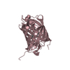
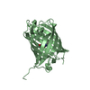
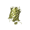
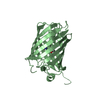
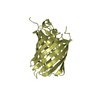
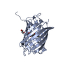
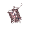
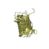
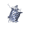
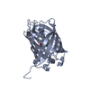
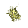
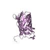
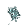
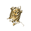
A: Far-red fluorescent protein mKate B: Far-red fluorescent protein mKate C: Far-red fluorescent protein mKate D: Far-red fluorescent protein mKate E: Far-red fluorescent protein mKate F: Far-red fluorescent protein mKate G: Far-red fluorescent protein mKate H: Far-red fluorescent protein mKate
Protocol: SINGLE WAVELENGTH / Monochromatic (M) / Laue (L): M / Scattering type: x-ray
Radiation wavelength
Wavelength: 1 Å / Relative weight: 1
Reflection
Resolution: 2.6→30 Å / Num. obs: 55727 / % possible obs: 99.9 %
Reflection shell
Resolution: 2.6→2.69 Å / Rmerge(I) obs: 0.506 / % possible all: 99.4
-
Processing
Software
Name
Version
Classification
NB
PHENIX
refinement
SCALEPACK
datascaling
REFMAC
refinement
PDB_EXTRACT
3.004
dataextraction
SERGUI
datacollection
HKL-2000
datareduction
HKL-2000
datascaling
MOLREP
phasing
DENZO
datareduction
Refinement
Resolution: 2.6→29.84 Å / Cor.coef. Fo:Fc: 0.945 / Cor.coef. Fo:Fc free: 0.88 / SU B: 13.087 / SU ML: 0.284 / ESU R Free: 0.394 / Stereochemistry target values: MAXIMUM LIKELIHOOD / Details: HYDROGENS HAVE BEEN ADDED IN THE RIDING POSITIONS
Rfactor
Num. reflection
% reflection
Rfree
0.263
2814
5.05 %
Rwork
0.17
-
-
obs
-
55727
99.7 %
Solvent computation
Bsol: 28.7 Å2 / ksol: 0.32 e/Å3
Displacement parameters
Biso mean: 44.52 Å2
Baniso -1
Baniso -2
Baniso -3
1-
2.394 Å2
0 Å2
-4.942 Å2
2-
-
0.706 Å2
0 Å2
3-
-
-
-3.1 Å2
Refinement step
Cycle: LAST / Resolution: 2.6→29.84 Å
Protein
Nucleic acid
Ligand
Solvent
Total
Num. atoms
14264
0
0
351
14615
Refine LS restraints
Refine-ID
Type
Dev ideal
X-RAY DIFFRACTION
f_bond_d
0.008
X-RAY DIFFRACTION
f_angle_d
0.935
X-RAY DIFFRACTION
f_dihedral_angle_d
13.204
X-RAY DIFFRACTION
f_chiral_restr
0.066
X-RAY DIFFRACTION
f_plane_restr
0.003
LS refinement shell
Resolution: 2.6→2.61 Å
Rfactor
Num. reflection
% reflection
Rwork
0.224
497
-
obs
-
-
93 %
+
About Yorodumi
-
News
-
Feb 9, 2022. New format data for meta-information of EMDB entries
New format data for meta-information of EMDB entries
Version 3 of the EMDB header file is now the official format.
The previous official version 1.9 will be removed from the archive.
In the structure databanks used in Yorodumi, some data are registered as the other names, "COVID-19 virus" and "2019-nCoV". Here are the details of the virus and the list of structure data.
Jan 31, 2019. EMDB accession codes are about to change! (news from PDBe EMDB page)
EMDB accession codes are about to change! (news from PDBe EMDB page)
The allocation of 4 digits for EMDB accession codes will soon come to an end. Whilst these codes will remain in use, new EMDB accession codes will include an additional digit and will expand incrementally as the available range of codes is exhausted. The current 4-digit format prefixed with “EMD-” (i.e. EMD-XXXX) will advance to a 5-digit format (i.e. EMD-XXXXX), and so on. It is currently estimated that the 4-digit codes will be depleted around Spring 2019, at which point the 5-digit format will come into force.
The EM Navigator/Yorodumi systems omit the EMD- prefix.
Related info.:Q: What is EMD? / ID/Accession-code notation in Yorodumi/EM Navigator
Yorodumi is a browser for structure data from EMDB, PDB, SASBDB, etc.
This page is also the successor to EM Navigator detail page, and also detail information page/front-end page for Omokage search.
The word "yorodu" (or yorozu) is an old Japanese word meaning "ten thousand". "mi" (miru) is to see.
Related info.:EMDB / PDB / SASBDB / Comparison of 3 databanks / Yorodumi Search / Aug 31, 2016. New EM Navigator & Yorodumi / Yorodumi Papers / Jmol/JSmol / Function and homology information / Changes in new EM Navigator and Yorodumi
 Movie
Movie Controller

 Yorodumi
 Open data
 Basic information
 Components
 Keywords
 Function and homology information
 Entacmaea quadricolor (sea anemone)
 X-RAY DIFFRACTION /
  Authors
 Citation
 Structure visualization
 Downloads & links
 Other downloads
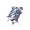


 PDBj
PDBj Assembly
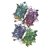


 Mass: 18.015 Da / Num. of mol.: 351 / Source method: isolated from a natural source / Formula: H2O
 Sample preparation
 / Beamline: 22-ID / Wavelength: 1 Å
 Processing