 Movie
Movie Controller

[English] 日本語
 Yorodumi
- PDB-2bh1: X-ray structure of the general secretion pathway complex of the N... -
+ Open data

- Basic information
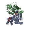
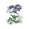
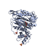
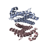
| Entry | Database: PDB / ID: 2bh1 | ||||||
|---|---|---|---|---|---|---|---|
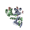
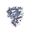
| Title | X-ray structure of the general secretion pathway complex of the N- terminal domain of EpsE and the cytosolic domain of EpsL of Vibrio cholerae | ||||||
 Components Components |
| ||||||
 Keywords Keywords | TRANSPORT PROTEIN / TYPE II SECRETION / VIBRIO CHOLERAE / EPS / GSP / TRANSMEMBRANE / TRANSPORT / ATP-BINDING | ||||||
| Function / homology |  Function and homology information Function and homology informationprotein-exporting ATPase activity / protein-secreting ATPase / Gram-negative-bacterium-type cell wall / protein secretion by the type II secretion system / type II protein secretion system complex / ATP hydrolysis activity / ATP binding / metal ion binding / plasma membrane Similarity search - Function | ||||||
| Biological species |   VIBRIO CHOLERAE (bacteria) VIBRIO CHOLERAE (bacteria) | ||||||
| Method |  X-RAY DIFFRACTION / SYNCHROTRON / MOLECULAR REPLACEMENT / Resolution: 2.4 Å X-RAY DIFFRACTION / SYNCHROTRON / MOLECULAR REPLACEMENT / Resolution: 2.4 Å | ||||||
 Authors Authors | Abendroth, J. / Murphy, P.M. / Mushtaq, A. / Bagdasarian, M. / Sandkvist, M. / Hol, W.G.J. | ||||||
 Citation Citation | Journal: J.Mol.Biol. / Year: 2005 Title: The X-Ray Structure of the Type II Secretion System Complex Formed by the N-Terminal Domain of Epse and the Cytoplasmic Domain of Epsl of Vibrio Cholerae Authors: Abendroth, J. / Murphy, P.M. / Mushtaq, A. / Bagdasarian, M. / Sandkvist, M. / Hol, W.G.J. | ||||||
| History |
| ||||||
| Remark 650 | HELIX DETERMINATION METHOD: AUTHOR PROVIDED. |
- Structure visualization
| Structure viewer | Molecule: MolmilJmol/JSmol |
|---|
- Downloads & links
-Download
| PDBx/mmCIF format | 2bh1.cif.gz | 135.2 KB | Display | PDBx/mmCIF format |
|---|---|---|---|---|
| PDB format | pdb2bh1.ent.gz | 104.7 KB | Display | PDB format |
| PDBx/mmJSON format | 2bh1.json.gz | Tree view | PDBx/mmJSON format | |
| Others |  Other downloads Other downloads |
-Validation report
| Arichive directory | https://data.pdbj.org/pub/pdb/validation_reports/bh/2bh1ftp://data.pdbj.org/pub/pdb/validation_reports/bh/2bh1 | HTTPS FTP |
|---|
-Related structure data
| Related structure data |  1yf5C  1w97S S: Starting model for refinement C: citing same article ( |
|---|---|
| Similar structure data |


-Links
 PDBj
PDBj
- Assembly
| Deposited unit | 
| ||||||||||||||||||||||||||||||||||||||||||||||||||||||||||||||||||||||||||||||||
|---|---|---|---|---|---|---|---|---|---|---|---|---|---|---|---|---|---|---|---|---|---|---|---|---|---|---|---|---|---|---|---|---|---|---|---|---|---|---|---|---|---|---|---|---|---|---|---|---|---|---|---|---|---|---|---|---|---|---|---|---|---|---|---|---|---|---|---|---|---|---|---|---|---|---|---|---|---|---|---|---|---|
| 1 | 
| ||||||||||||||||||||||||||||||||||||||||||||||||||||||||||||||||||||||||||||||||
| 2 | 
| ||||||||||||||||||||||||||||||||||||||||||||||||||||||||||||||||||||||||||||||||
| Unit cell |
| ||||||||||||||||||||||||||||||||||||||||||||||||||||||||||||||||||||||||||||||||
| Noncrystallographic symmetry (NCS) | NCS domain:
NCS domain segments: Component-ID: 1 / Refine code: 6
NCS ensembles :
NCS oper:
| ||||||||||||||||||||||||||||||||||||||||||||||||||||||||||||||||||||||||||||||||
| Details | FOR THE HETERO_ASSEMBLY DESCRIBED BY REMARK 350 |
-Components
| #1: Protein | Mass: 27983.732 Da / Num. of mol.: 2 / Fragment: CYTOPLASMIC DOMAIN, RESIDUES 5-246 Source method: isolated from a genetically manipulated source Source: (gene. exp.) VIBRIO CHOLERAE (bacteria) / Production host: #2: Protein | Mass: 11187.723 Da / Num. of mol.: 2 / Fragment: DOMAIN N1, RESIDUES 1-96 Source method: isolated from a genetically manipulated source Source: (gene. exp.) VIBRIO CHOLERAE (bacteria) / Production host: #3: Chemical | ChemComp-CA / |   Mass: 40.078 Da / Num. of mol.: 1 / Source method: obtained synthetically / Formula: Ca Mass: 40.078 Da / Num. of mol.: 1 / Source method: obtained synthetically / Formula: Ca#4: Water | ChemComp-HOH / |  Mass: 18.015 Da / Num. of mol.: 177 / Source method: isolated from a natural source / Formula: H2O Mass: 18.015 Da / Num. of mol.: 177 / Source method: isolated from a natural source / Formula: H2O |
|---|
-Experimental details
-Experiment
| Experiment | Method: X-RAY DIFFRACTION / Number of used crystals: 1 |
|---|
- Sample preparation
| Crystal | Density Matthews: 2.3 Å3/Da / Density % sol: 45 % |
|---|---|
| Crystal grow | pH: 5.5 Details: 200MM CAOAC2, 100MM BISTRIS PH5.5, 25% PEG 3350, pH 5.50 |
-Data collection
| Diffraction | Mean temperature: 100 K |
|---|---|
| Diffraction source | Source: SYNCHROTRON / Site: ALS  / Beamline: 8.2.2 / Wavelength: 0.9999 / Beamline: 8.2.2 / Wavelength: 0.9999 |
| Detector | Type: ADSC CCD / Detector: CCD / Date: Sep 17, 2004 |
| Radiation | Protocol: SINGLE WAVELENGTH / Monochromatic (M) / Laue (L): M / Scattering type: x-ray |
| Radiation wavelength | Wavelength: 0.9999 Å / Relative weight: 1 |
| Reflection | Resolution: 2.4→20 Å / Num. obs: 27927 / % possible obs: 99.8 % / Redundancy: 3.6 % / Rmerge(I) obs: 0.07 / Net I/σ(I): 10.7 |
| Reflection shell | Resolution: 2.4→2.5 Å / Redundancy: 3.6 % / Rmerge(I) obs: 0.69 / Mean I/σ(I) obs: 1.9 / % possible all: 100 |
- Processing
| Software |
| ||||||||||||||||||||||||||||||||||||||||||||||||||||||||||||||||||||||||||||||||||||||||||||||||||||||||||||||||||||||||||||||||||||||||||||||||||||||||||||||||||||||||||||||||||||||
|---|---|---|---|---|---|---|---|---|---|---|---|---|---|---|---|---|---|---|---|---|---|---|---|---|---|---|---|---|---|---|---|---|---|---|---|---|---|---|---|---|---|---|---|---|---|---|---|---|---|---|---|---|---|---|---|---|---|---|---|---|---|---|---|---|---|---|---|---|---|---|---|---|---|---|---|---|---|---|---|---|---|---|---|---|---|---|---|---|---|---|---|---|---|---|---|---|---|---|---|---|---|---|---|---|---|---|---|---|---|---|---|---|---|---|---|---|---|---|---|---|---|---|---|---|---|---|---|---|---|---|---|---|---|---|---|---|---|---|---|---|---|---|---|---|---|---|---|---|---|---|---|---|---|---|---|---|---|---|---|---|---|---|---|---|---|---|---|---|---|---|---|---|---|---|---|---|---|---|---|---|---|---|---|
| Refinement | Method to determine structure: MOLECULAR REPLACEMENT Starting model: PDB ENTRY 1W97 Resolution: 2.4→20 Å / Cor.coef. Fo:Fc: 0.962 / Cor.coef. Fo:Fc free: 0.93 / SU B: 21.652 / SU ML: 0.253 / TLS residual ADP flag: LIKELY RESIDUAL / Cross valid method: THROUGHOUT / ESU R: 0.412 / ESU R Free: 0.268 / Stereochemistry target values: MAXIMUM LIKELIHOOD / Details: HYDROGENS HAVE BEEN ADDED IN THE RIDING POSITIONS.
| ||||||||||||||||||||||||||||||||||||||||||||||||||||||||||||||||||||||||||||||||||||||||||||||||||||||||||||||||||||||||||||||||||||||||||||||||||||||||||||||||||||||||||||||||||||||
| Solvent computation | Ion probe radii: 0.8 Å / Shrinkage radii: 0.8 Å / VDW probe radii: 1.2 Å / Solvent model: MASK | ||||||||||||||||||||||||||||||||||||||||||||||||||||||||||||||||||||||||||||||||||||||||||||||||||||||||||||||||||||||||||||||||||||||||||||||||||||||||||||||||||||||||||||||||||||||
| Displacement parameters | Biso mean: 50.05 Å2
| ||||||||||||||||||||||||||||||||||||||||||||||||||||||||||||||||||||||||||||||||||||||||||||||||||||||||||||||||||||||||||||||||||||||||||||||||||||||||||||||||||||||||||||||||||||||
| Refinement step | Cycle: LAST / Resolution: 2.4→20 Å
| ||||||||||||||||||||||||||||||||||||||||||||||||||||||||||||||||||||||||||||||||||||||||||||||||||||||||||||||||||||||||||||||||||||||||||||||||||||||||||||||||||||||||||||||||||||||
| Refine LS restraints |
|