 Movie
Movie Controller

[English] 日本語
 Yorodumi
- PDB-1yf5: Cyto-Epsl: The Cytoplasmic Domain Of Epsl, An Inner Membrane Comp... -
+ Open data

- Basic information
| Entry | Database: PDB / ID: 1yf5 | ||||||
|---|---|---|---|---|---|---|---|
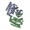
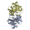
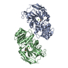
| Title | Cyto-Epsl: The Cytoplasmic Domain Of Epsl, An Inner Membrane Component Of The Type II Secretion System Of Vibrio Cholerae | ||||||
 Components Components | General secretion pathway protein L | ||||||
 Keywords Keywords | TRANSPORT PROTEIN / Type II secretion / secretory protein | ||||||
| Function / homology |  Function and homology information Function and homology informationGram-negative-bacterium-type cell wall / protein secretion by the type II secretion system / type II protein secretion system complex / plasma membrane Similarity search - Function | ||||||
| Biological species |   Vibrio cholerae (bacteria) Vibrio cholerae (bacteria) | ||||||
| Method |  X-RAY DIFFRACTION / SYNCHROTRON / MOLECULAR REPLACEMENT / Resolution: 2.75 Å X-RAY DIFFRACTION / SYNCHROTRON / MOLECULAR REPLACEMENT / Resolution: 2.75 Å | ||||||
 Authors Authors | Abendroth, J. / Murphy, P. / Mushtaq, A. / Sandkvist, M. / Bagdasarian, M. / Hol, W.G. | ||||||
 Citation Citation | Journal: J.Mol.Biol. / Year: 2005 Title: The X-ray Structure of the Type II Secretion System Complex Formed by the N-terminal Domain of EpsE and the Cytoplasmic Domain of EpsL of Vibrio cholerae. Authors: Abendroth, J. / Murphy, P. / Sandkvist, M. / Bagdasarian, M. / Hol, W.G. #1: Journal: J.Mol.Biol. / Year: 2004Title: The Structure of the Cytoplasmic Domain of Epsl, an Inner Membrane Component of the Type II Secretio System of Vibrio Cholerae: An Unusual Member of the Actin-Like ATPase Superfamily Authors: Abendroth, J. / Bagdasarian, M. / Sandkvist, M. / Hol, W.G. | ||||||
| History |
|
- Structure visualization
| Structure viewer | Molecule: MolmilJmol/JSmol |
|---|
- Downloads & links
-Download
| PDBx/mmCIF format | 1yf5.cif.gz | 59.3 KB | Display | PDBx/mmCIF format |
|---|---|---|---|---|
| PDB format | pdb1yf5.ent.gz | 42.8 KB | Display | PDB format |
| PDBx/mmJSON format | 1yf5.json.gz | Tree view | PDBx/mmJSON format | |
| Others |  Other downloads Other downloads |
-Validation report
| Arichive directory | https://data.pdbj.org/pub/pdb/validation_reports/yf/1yf5ftp://data.pdbj.org/pub/pdb/validation_reports/yf/1yf5 | HTTPS FTP |
|---|
-Related structure data
| Related structure data |  2bh1C  1w97S S: Starting model for refinement C: citing same article ( |
|---|---|
| Similar structure data |


-Links
 PDBj
PDBj- Assembly
| Deposited unit | 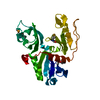
| ||||||||
|---|---|---|---|---|---|---|---|---|---|
| 1 | 
| ||||||||
| Unit cell |
| ||||||||
| Details | the second half of the dimer is generated by the crystallographic two-fold: -x+2, y, -z |
-Components
| #1: Protein | Mass: 28528.854 Da / Num. of mol.: 1 Source method: isolated from a genetically manipulated source Source: (gene. exp.) Vibrio cholerae (bacteria) / Gene: epsL / Production host: |
|---|---|
| #2: Water | ChemComp-HOH /  Mass: 18.015 Da / Num. of mol.: 9 / Source method: isolated from a natural source / Formula: H2O Mass: 18.015 Da / Num. of mol.: 9 / Source method: isolated from a natural source / Formula: H2O |
| Has protein modification | Y |
-Experimental details
-Experiment
| Experiment | Method: X-RAY DIFFRACTION / Number of used crystals: 1 |
|---|
- Sample preparation
| Crystal | Density Matthews: 2.5 Å3/Da / Density % sol: 51.2 % |
|---|---|
| Crystal grow | Temperature: 293 K / Method: vapor diffusion, sitting drop / pH: 8 Details: 15% PEG 6000, 150mM calcium acetate, 100mM tris pH 8, VAPOR DIFFUSION, SITTING DROP, temperature 293K |
-Data collection
| Diffraction | Mean temperature: 100 K |
|---|---|
| Diffraction source | Source: SYNCHROTRON / Site: APS  / Beamline: 19-ID / Wavelength: 0.97951 Å / Beamline: 19-ID / Wavelength: 0.97951 Å |
| Detector | Type: ADSC QUANTUM 315 / Detector: CCD / Date: Mar 29, 2003 |
| Radiation | Protocol: SINGLE WAVELENGTH / Monochromatic (M) / Laue (L): M / Scattering type: x-ray |
| Radiation wavelength | Wavelength: 0.97951 Å / Relative weight: 1 |
| Reflection | Resolution: 2.74→20 Å / Num. all: 7122 / Num. obs: 7122 / % possible obs: 95.8 % / Observed criterion σ(F): 0 / Observed criterion σ(I): 0 / Redundancy: 7 % / Biso Wilson estimate: 87.4 Å2 / Rmerge(I) obs: 0.08 / Net I/σ(I): 15.1 |
| Reflection shell | Resolution: 2.74→2.8 Å / Redundancy: 5.2 % / Rmerge(I) obs: 0.37 / Mean I/σ(I) obs: 3.4 / Rsym value: 0.37 / % possible all: 81 |
- Processing
| Software |
| ||||||||||||||||||||||||||||||||||||||||||||||||||||||||||||||||||||||||||||||||||||||||||
|---|---|---|---|---|---|---|---|---|---|---|---|---|---|---|---|---|---|---|---|---|---|---|---|---|---|---|---|---|---|---|---|---|---|---|---|---|---|---|---|---|---|---|---|---|---|---|---|---|---|---|---|---|---|---|---|---|---|---|---|---|---|---|---|---|---|---|---|---|---|---|---|---|---|---|---|---|---|---|---|---|---|---|---|---|---|---|---|---|---|---|---|
| Refinement | Method to determine structure: MOLECULAR REPLACEMENT Starting model: 1w97 Resolution: 2.75→20 Å / Cor.coef. Fo:Fc: 0.953 / Cor.coef. Fo:Fc free: 0.919 / SU B: 42.646 / SU ML: 0.35 / TLS residual ADP flag: LIKELY RESIDUAL / Cross valid method: THROUGHOUT / σ(F): 0 / ESU R Free: 0.402 / Stereochemistry target values: MAXIMUM LIKELIHOOD / Details: HYDROGENS HAVE BEEN ADDED IN THE RIDING POSITIONS
| ||||||||||||||||||||||||||||||||||||||||||||||||||||||||||||||||||||||||||||||||||||||||||
| Solvent computation | Ion probe radii: 0.8 Å / Shrinkage radii: 0.8 Å / VDW probe radii: 1.2 Å / Solvent model: MASK | ||||||||||||||||||||||||||||||||||||||||||||||||||||||||||||||||||||||||||||||||||||||||||
| Displacement parameters | Biso mean: 76.835 Å2
| ||||||||||||||||||||||||||||||||||||||||||||||||||||||||||||||||||||||||||||||||||||||||||
| Refinement step | Cycle: LAST / Resolution: 2.75→20 Å
| ||||||||||||||||||||||||||||||||||||||||||||||||||||||||||||||||||||||||||||||||||||||||||
| Refine LS restraints |
| ||||||||||||||||||||||||||||||||||||||||||||||||||||||||||||||||||||||||||||||||||||||||||
| LS refinement shell | Resolution: 2.75→2.896 Å / Total num. of bins used: 10
| ||||||||||||||||||||||||||||||||||||||||||||||||||||||||||||||||||||||||||||||||||||||||||
| Refinement TLS params. | Method: refined / Origin x: 45.6585 Å / Origin y: -4.0897 Å / Origin z: 12.534 Å
| ||||||||||||||||||||||||||||||||||||||||||||||||||||||||||||||||||||||||||||||||||||||||||
| Refinement TLS group |
|
