 Movie
Movie Controller

[English] 日本語
 Yorodumi
- PDB-1s28: Crystal Structure of AvrPphF ORF1, the Chaperone for the Type III... -
+ Open data

- Basic information
| Entry | Database: PDB / ID: 1s28 | ||||||
|---|---|---|---|---|---|---|---|
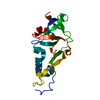
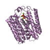
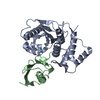
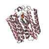
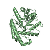
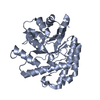
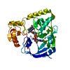
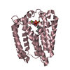
| Title | Crystal Structure of AvrPphF ORF1, the Chaperone for the Type III Effector AvrPphF ORF2 from P. syringae | ||||||
 Components Components | ORF1 | ||||||
 Keywords Keywords | CHAPERONE / Type III Chaperone | ||||||
| Function / homology | Tir chaperone protein (CesT) family / Tir chaperone protein (CesT) family / Yope Regulator; Chain: A, - #10 / Yope Regulator; Chain: A, / protein secretion by the type III secretion system / 2-Layer Sandwich / Alpha Beta / ORF1 Function and homology information Function and homology information | ||||||
| Biological species |  Pseudomonas syringae pv. phaseolicola (bacteria) Pseudomonas syringae pv. phaseolicola (bacteria) | ||||||
| Method |  X-RAY DIFFRACTION / SYNCHROTRON / MAD / Resolution: 3 Å X-RAY DIFFRACTION / SYNCHROTRON / MAD / Resolution: 3 Å | ||||||
 Authors Authors | Singer, A.U. / Desveaux, D. / Betts, L. / Chang, J.H. / Nimchuk, Z. / Grant, S.R. / Dangl, J.L. / Sondek, J. | ||||||
 Citation Citation | Journal: Structure / Year: 2004 Title: Crystal Structures of the Type III Effector Protein AvrPphF and Its Chaperone Reveal Residues Required for Plant Pathogenesis Authors: Singer, A.U. / Desveaux, D. / Betts, L. / Chang, J.H. / Nimchuk, Z. / Grant, S.R. / Dangl, J.L. / Sondek, J. | ||||||
| History |
| ||||||
| Remark 999 | sequence The last five residues in C-terminus vary from the published sequence, which authors ... sequence The last five residues in C-terminus vary from the published sequence, which authors believe may have a sequencing error. |
- Structure visualization
| Structure viewer | Molecule: MolmilJmol/JSmol |
|---|
- Downloads & links
-Download
| PDBx/mmCIF format | 1s28.cif.gz | 115.8 KB | Display | PDBx/mmCIF format |
|---|---|---|---|---|
| PDB format | pdb1s28.ent.gz | 92.3 KB | Display | PDB format |
| PDBx/mmJSON format | 1s28.json.gz | Tree view | PDBx/mmJSON format | |
| Others |  Other downloads Other downloads |
-Validation report
| Arichive directory | https://data.pdbj.org/pub/pdb/validation_reports/s2/1s28ftp://data.pdbj.org/pub/pdb/validation_reports/s2/1s28 | HTTPS FTP |
|---|
-Related structure data


-Links
 PDBj
PDBj
- Assembly
| Deposited unit | 
| ||||||||
|---|---|---|---|---|---|---|---|---|---|
| 1 | 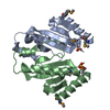
| ||||||||
| 2 | 
| ||||||||
| 3 |
| ||||||||
| Unit cell |
| ||||||||
| Details | The molecule is a biological dimer, though there were 4 molecules in the asymmetric unit. Molecule 2 can superimpose on molecule 1 with the transformation matrix: -0.985 -0.173 0.003 146.58 -0.173 0.985 -0.016 12.77 0 -0.016 -1.0 3.38 |
-Components
| #1: Protein | Mass: 15489.323 Da / Num. of mol.: 4 / Fragment: AvrPphf ORF1 Source method: isolated from a genetically manipulated source Source: (gene. exp.) Pseudomonas syringae pv. phaseolicola (bacteria)Species: Pseudomonas savastanoi / Gene: AvrPphF ORF1 / Plasmid: pProEX-HTa / Production host: #2: Chemical | ChemComp-SO4 /   Mass: 96.063 Da / Num. of mol.: 4 / Source method: obtained synthetically / Formula: SO4 Mass: 96.063 Da / Num. of mol.: 4 / Source method: obtained synthetically / Formula: SO4Has protein modification | Y | |
|---|
-Experimental details
-Experiment
| Experiment | Method: X-RAY DIFFRACTION / Number of used crystals: 1 |
|---|
- Sample preparation
| Crystal | Density Matthews: 3.85 Å3/Da / Density % sol: 67.82 % |
|---|---|
| Crystal grow | Temperature: 277 K / Method: vapor diffusion, sitting drop / pH: 7.5 Details: Ammonium Sulfate,Tris 7.5, VAPOR DIFFUSION, SITTING DROP, temperature 277K |
-Data collection
| Diffraction | Mean temperature: 100 K | ||||||||||||
|---|---|---|---|---|---|---|---|---|---|---|---|---|---|
| Diffraction source | Source: SYNCHROTRON / Site: APS  / Beamline: 22-ID / Wavelength: 0.97925, 0.97936, 0.97167 / Beamline: 22-ID / Wavelength: 0.97925, 0.97936, 0.97167 | ||||||||||||
| Detector | Type: MARRESEARCH / Detector: CCD / Date: Apr 23, 2003 / Details: vertically focussing mirror | ||||||||||||
| Radiation | Monochromator: Double Crystal Monochromater Si-220 / Protocol: MAD / Monochromatic (M) / Laue (L): M / Scattering type: x-ray | ||||||||||||
| Radiation wavelength |
| ||||||||||||
| Reflection | Resolution: 3→20 Å / Num. all: 18977 / Num. obs: 18973 / % possible obs: 100 % / Observed criterion σ(F): 0 / Observed criterion σ(I): -3 / Redundancy: 7.4 % / Biso Wilson estimate: 72.7 Å2 / Rmerge(I) obs: 0.096 / Net I/σ(I): 26.9 | ||||||||||||
| Reflection shell | Resolution: 3→3.11 Å / Redundancy: 7.5 % / Rmerge(I) obs: 0.257 / Mean I/σ(I) obs: 7.66 / Num. unique all: 1851 / % possible all: 100 |
- Processing
| Software |
| ||||||||||||||||||||||||||||||||||||
|---|---|---|---|---|---|---|---|---|---|---|---|---|---|---|---|---|---|---|---|---|---|---|---|---|---|---|---|---|---|---|---|---|---|---|---|---|---|
| Refinement | Method to determine structure: MAD / Resolution: 3→19.97 Å / Rfactor Rfree error: 0.01 / Data cutoff high absF: 1713413.02 / Data cutoff low absF: 0 / Isotropic thermal model: RESTRAINED / Cross valid method: THROUGHOUT / σ(F): 1.5 / Stereochemistry target values: Engh & Huber
| ||||||||||||||||||||||||||||||||||||
| Solvent computation | Solvent model: FLAT MODEL / Bsol: 10 Å2 / ksol: 0.280684 e/Å3 | ||||||||||||||||||||||||||||||||||||
| Displacement parameters | Biso mean: 43.2 Å2
| ||||||||||||||||||||||||||||||||||||
| Refine analyze |
| ||||||||||||||||||||||||||||||||||||
| Refinement step | Cycle: LAST / Resolution: 3→19.97 Å
| ||||||||||||||||||||||||||||||||||||
| Refine LS restraints |
| ||||||||||||||||||||||||||||||||||||
| Refine LS restraints NCS | NCS model details: CONSTR | ||||||||||||||||||||||||||||||||||||
| LS refinement shell | Resolution: 3→3.19 Å / Rfactor Rfree error: 0.028 / Total num. of bins used: 6
| ||||||||||||||||||||||||||||||||||||
| Xplor file |
|
